This is an automatically translated article.
The article is professionally consulted by Master, Doctor Nguyen Quang Duc - Doctor of Nuclear Medicine - Department of Diagnostic Imaging and Nuclear Medicine - Vinmec Times City International General Hospital.In applied medicine and what is the role of radioisotopes? Currently, the application of radioisotopes in the diagnosis and treatment of cancer diseases is becoming popular and widespread, thanks to its effectiveness, short duration and safety.
1. What is a radioactive isotope?
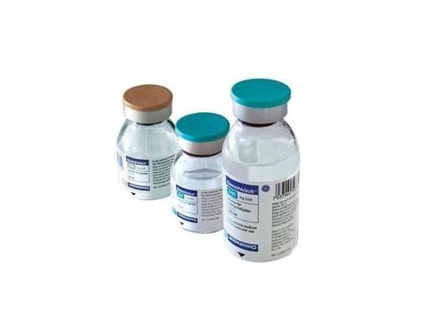
What is a radioactive isotope? These are isotopes of an element, discovered in 1930. In nuclear medicine, radioisotopes are used for diagnostic purposes based on the principle that radioactive isotopes enter the body in the form of radioactive isotopes. Radiopharmaceuticals and participate in physiological processes do not cause pharmacological effects.Radiopharmaceuticals, also known as radiopharmaceuticals, include radioactive isotopes attached to a carrier that can be taken orally or injected into the body. Depending on the purpose and the organ that needs to be examined and diagnosed, different carriers will be used.
2. Radioisotope applications in diagnosis
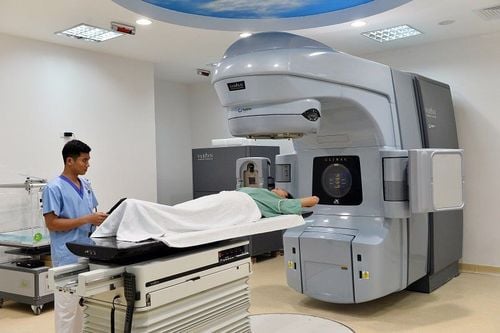
2.1 Devices for application of radioisotopes in diagnostics Radioisotope applications in diagnostics is a technique of using a scanner to measure radioactivity from the outside and to record images of the distribution of isotopes. radioactive taste when they are inside the body.The following types of scanners are available:
Scanner: Linear scanner with a portable detector Gamma camera: A scanner with a large and non-portable probe that can cover the whole body to measure radioactivity of the whole body or of an area. SPECT (Single Photon Emission Computed Tomography): A photon emission tomography scanner is similar to a gamma camera but can rotate 360 degrees. Currently, SPECT is a diagnostic radioisotope application device that is widely used in the examination and treatment of neurological and cardiovascular diseases, determining the stage of the disease and evaluating the effectiveness of cancer treatment, .. PET (Positron Emission Tomography): The positron emission tomography machine is like the SPECT machine but records by measuring positron emission. SPECT/CT, PET/CT : A combination of SPECT, PET and CT machines to obtain both functional and anatomical images.

Active transport mechanism: Difference in the concentration distribution of some substances that always exist in living organisms, based on which substances can be transported from places of low concentration to high concentration. Diffusion: The application of radioisotopes in diagnosis is based on the mechanism of some organs in the body, when the outer barrier is damaged, it will create conditions for radioactive isotopes to attach to the carrier and travel with the blood. diffuse into the affected area. Metabolism: In the body, a tumor is a collection of cells that consume more glucose. When radioactive isotopes in the form of organic or inorganic salts are introduced into the body and participate in the metabolism, they will be more concentrated in the damaged areas. Deposition: Depending on the examination agency, the radioisotope application in diagnosis will be selected and attached to a suitable colloidal carrier so that when entering the body, it can be deposited to facilitate diagnostic imaging. . Elimination: In contrast to the deposition mechanism, some organs such as the liver and kidneys have the function of elimination to be diagnosed, requiring radioactive waste to enter. Application of radioisotopes in diagnosis, in addition to the above-mentioned mechanisms, there are a number of other mechanisms to deliver radiopharmaceuticals to the target organs to be examined, such as phagocytosis, immunity, temporary microvascular occlusion. time, specific receivers... To ensure that the target - no target ratio reaches the minimum value for diagnosis, it is necessary to pay attention to the causes of this decrease, especially the patient's body.

Thyroid scan : Thyroid scan is performed to locate and evaluate the shape and size of the thyroid gland; provide functional imaging of thyroid nodules; monitoring and evaluation of functional status before and after thyroid cancer surgery; helps to differentiate the diagnosis from a cervical-mediastinal tumor. Myocardial perfusion scintigraphy: The application of radioisotopes in the diagnosis of myocardial perfusion scintigraphy is based on the principle that some radioisotopes will remain in the myocardium for a while and emit gamma radiation for recording. Figure. Areas of hypoperfusion or radiation defect are areas of poor or no perfusion. Myocardial perfusion scintigraphy needs to be performed when the body is in a state of exercise and rest in order to accurately assess the myocardial blood supply as well as the viability and regional functioning status of the myocardium. . Renal scintigraphy: The application of radioisotopes in renal scintigraphy must select radioisotopes emitting gamma radiation. These drugs are quickly absorbed by the kidneys and then participate in the process of filtration - elimination. However, the body stores these drugs for a long enough time that the scanner can measure and record the distribution of radioactivity in the kidney. Brain scan: When the blood-brain barrier is damaged (due to diseases such as cerebral ischemia, brain abscess, brain cancer or trauma, ...) will create conditions for radioactive isotopes to attach to the carrier. from the blood to the affected area. The application of radioisotopes in brain scintigraphy will record highly concentrated radioactive images in the outer space when the blood-brain barrier is damaged, which is the hot area; or areas of reduction or defect in the occurrence of lesions. Bone scan: Bone scan is performed in cases such as fracture, bone cancer, osteomyelitis, bone metastatic cancer, ... to look for areas of bone proliferation, based on the mechanism of increased perfusion. blood, cell proliferation, increased metabolism in areas of pathological lesions, which are areas of high radiation concentration. Radioisotopes in diagnosis are used to detect early pathological lesions in the brain, thyroid gland, heart muscle, kidneys, bones, and help monitor and evaluate treatment.
Currently, Vinmec International General Hospital has become a leading prestigious address in disease screening with modern techniques, proud to be the first private hospital in Vietnam to deploy this technique. Modern and accurate diagnosis with the most modern 128-sequence PET/CT system in Southeast Asia, for accurate images and short imaging time, the team of doctors are leading experts with high expertise and rich experience. Experience creates trust for customers when experiencing medical examination and treatment services at Vinmec.
If you have a need for consultation and examination at Vinmec Hospitals under the nationwide health system, please book an appointment on the website for service.
Please dial HOTLINE for more information or register for an appointment HERE. Download MyVinmec app to make appointments faster and to manage your bookings easily.


