This is an automatically translated article.
Posted by Doctor Nguyen Duc Tho - Department of Surgical Anesthesia - Vinmec Central Park International Hospital
Placing femoral nerve catheters to relieve pain after lower extremity surgery is a new technique with many advantages when not using Morphine and morphine precursors, helping to reduce the side effects of morphine such as nausea and vomiting. , vomiting, urinary retention, constipation, even addictive.
1. What is catheterization, why should catheters be placed?
Anesthesia under ultrasound guidance is a method of injecting anesthetic into the nerve sheath close to the nerves to help relieve pain during surgery and reduce pain after surgery.
Because the effect of local anesthetic is usually short, in order to relieve pain for a long time for the patient after surgery, the doctor must thread a plastic catheter (catheter) into the nerve sheath to continuously or intermittently pump the anesthetic. close to the nerve without the need for repeated anesthetic injections. Cathter can be saved 2 to 3 days after surgery to reduce patient pain
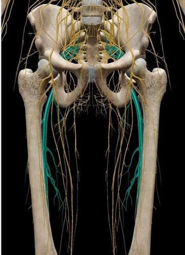
2. Femoral Nerve Anatomy
Is the largest branch of the lumbar plexus, formed by the posterior branches of the lumbar nerves LII, II, IV. Enters the groove of the LS muscle and then passes below the midpoint of the inguinal ligament (in the lumbosacral capsule) down to the femoral triangle lateral to the artery and divides into 3 types of branches. Muscle branches: works for the comb muscle, the muscle, the quadriceps muscle of the thigh, a part of the long adductor muscle. Dermis branches: poke through the sensory muscle for the front of the thigh. The saphenous nerve: passes through the femoral triangle into the adductor duct, crosses anteriorly to the artery, and then protrudes superficially and divides into two branches: The inferior patellar branch feels the facial skin in the knee. Medial dermis branch: sensation of the facial skin in the lower leg and part of the heel.

3. What is Catheter?
Catheter is a soft plastic catheter specially manufactured for medical use, they are inserted into internal body cavities such as blood vessels, nerve sheaths, pleural space, abdominal cavity, bladder, for the purpose of pumping drugs, providing fluids to the body, or draining body fluids out. This catheter can be temporarily left in the body until targeted therapy is achieved.
4. Designation
The surgery of the front of the thigh, surgery of the knee area (combination of the kneecap, endoscopic removal of the meniscus of the knee joint, reconstruction of the knee ligament). Frontal surgery in the lower leg. However, the anesthesiologist can assess the extent and area of surgery pain to decide whether to use this method or combine it with other methods to reduce pain or not.
5. Contraindications
Patient disagrees. The patient is allergic to the anesthetic. Infection of the anesthetic area. Sepsis. Epilepsy and psychiatric patients. Had surgery in the area of anesthesia. Massive blood loss.. Severe hypotension. Patients with coagulopathy or on anticoagulants. Uncooperative patient (may be combined with anesthesia).
6. Implementation
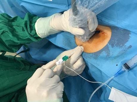
The patient lies supine with the thighs turned outward. The anesthesiologist will use an ultrasound machine to locate the femoral nerve, then use a specialized needle under ultrasound guidance, insert the needle into the connective capsule close to the nerve, and then insert the tube. catheter (catheter) through the needle into the appropriate position inside the capsule adjacent to the nerve.
Placement of the femoral nerve catheter can be performed in 2 positions, the femoral arch (horizontal inguinal crease) or in the adductor canal (medial third of the mid-thigh) depending on the surgical site and the area to be anesthetized
Wire The large femoral nerve is located in larger anatomical compartments, so a large volume of local anesthetic is required. This means that precise standards for drug volume and total dose are required to avoid overdosage of local anesthetic and the risk of toxicity.

7. Accidents and complications
Because the femoral nerve is quite large, moreover, under the guidance of ultrasound, doctors can easily determine the exact location of the nerve to insert the needle, so complications in femoral nerve anesthesia are often less. encounter
Possible complications are Nerve damage (a sensory disturbance in an area of the skin that is controlled by nerves). Poisoning by an overdose of local anesthetics or the introduction of local anesthetics into the blood vessels. Infection at the catheter site in case of long-term catheter retention.
8. Conclusion
With advances in technology, especially under ultrasound guidance, anesthesiologists are able to precisely identify the nerve to be anesthetized. Use a specialized needle for anesthesia to enter the nerve sheath, combine ultrasound and a nerve detector to identify the nerves located in the path of the needle, avoiding puncture of the blood vessel. As a result, complications and complications such as nerve damage, drug injection into the blood vessel, causing anesthetic toxicity, incorrect injection leading to technical failure are minimized.
Vinmec Hospital is currently applying this technique to relieve pain very effectively for patients after surgery, bringing satisfaction to patients.
Please dial HOTLINE for more information or register for an appointment HERE. Download MyVinmec app to make appointments faster and to manage your bookings easily.


