This is an automatically translated article.
Posted by Doctor Mai Vien Phuong - Department of Medical Examination & Internal Medicine - Vinmec Central Park International General Hospital.
Magnesium is required for many metabolic processes, but hypomagnesemia is common in hospitalized patients, especially in critically ill patients. Given the high morbidity and mortality reported in patients with hypotension, prompt diagnosis and treatment are of primary importance. This article will review magnesium basics including evaluation tests, causes of hypomagnesemia, and provide guidance on effective ways to treat magnesium deficiency.
1. Introduction to Magnesium
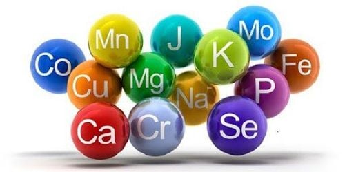
Magnesium (Mg), the fourth most abundant cation in the body, is a cofactor in more than 300 enzymatic reactions and plays an important role in protein, DNA and RNA synthesis. It is important for muscle contraction and relaxation, nerve function, heart rate, blood vessel tone, and bone formation. Ninety-nine percent of total Mg in the body is intracellular (bone, skeletal muscle, soft tissue) with only ~1% found in serum and red blood cells (extracellular). A large percentage (70-80%) of serum Mg is ionized (easily filtered by the kidneys), 20-30% bound to proteins (mainly albumin), and only ~ 1-2% complexed with anions .
Magnesium homeostasis is maintained by the intestines, bones and kidneys. It is absorbed most efficiently in the ileum with some absorption occurring in the colon via intracellular passivation and stored in bone; Excess magnesium is eliminated by the kidneys as well as in the feces. Only about 30-50% of the total dietary Mg consumed is absorbed in the intestine because other nutrients present in the gut (fiber, phytate, oxalate, phosphate) can bind cations and reduce the absorption of cations. its absorption. Magnesium status determines Mg absorption with more of this mineral being absorbed if Mg levels are low. The kidneys reabsorb ~95% of the filtered Mg and excrete only ~3 - 5% in the urine, unless rapid infusion is given.
2. Causes of hypomagnesemia:
Gastrointestinal lesions:Poor absorption (celiac, IBD) Small bowel bypass surgery, short bowel syndrome, gastrointestinal fistulae Diarrhea, vomiting, nasogastric aspiration Long-term use of pump inhibitors proton Excessive use of laxatives Primary intestinal hypoglycemia

Nôn mửa là một trong các nguyên nhân tại đường tiêu hóa gây hạ Magie trong máu
Reduced Mg intake:
Malnutrition, alcoholism Low magnesium diet Intravenous nutrition deficiency / low Mg Diabetic ketoacidosis Acute pancreatitis Renal magnesium loss:
Drug-induced renal magnesium loss Cyclic Diuretics & Thiazides Aminoglycoside Amphotericin Cyclosporine Tacrolimus Cisplatin Pentamidine Foscarnet Anti-EGFR Antibodies Renal Magnesium Loss Due to Polyuria Renal Magnesium Loss Due to Tubular Disorders Gitelman Syndrome Bartter Syndrome Diabetes
3. Condition assessment
Serum magnesium
There are several methods to assess Mg levels. The most common in clinical practice is the measurement of serum Mg. This test is widely available and inexpensive, but does not correlate with tissue magnesium stores. Serum Mg is a poor predictor of total body Mg as only 0.3% of total body Mg is found in serum.
Urine excretion test
The urine Mg excretion test is not commonly used in clinical practice, as it requires a 24-hour urine sample collection, which can be difficult to obtain. Renal Mg excretion follows circadian rhythms with the greatest amount of Mg excreted at night; Therefore, adequate 24-hour urine collection is essential for accurate assessment of absorption and excretion. High urinary excretion of Mg is a sign of kidney failure while low levels may indicate inadequate intake or absorption.

Bác sĩ có thể kiểm tra bài tiết nước tiểu đánh giá tình trạng hạ magie
“Magnesium retention test” or “load test” is a more sensitive indicator of Mg deficiency. It has been used to identify patients with suspected Mg deficiency during normal administration. If more than 60-70% of Mg is excreted in the urine following intravenous infusion, Mg deficiency is unlikely. This test, although more sensitive, can still give false positives or false negatives. Normal renal Mg processing is required for this test to be useful. Magnesium loss due to diabetes, medication or alcohol consumption can lead to false negative results, while with compromised kidney function one may see false positive results. The luminescence may be a confounding factor as older individuals tend to retain more Mg than younger patients. The Mg retention test should not be used in renal failure or transplant patients receiving cyclosporin or tacrolimus, both of which waste Mg in the urine.
4. Lowering Magnesium in the Blood
Hypomagnesemia is defined as a serum Mg concentration <1.8 mg/dL (normal range: 1.8 mg/dL - 2.3 mg/dL). Causes of hypomagnesaemia include poor oral intake, Mg deficiency from parenteral solution, altered absorption, and increased gastrointestinal loss in patients with diarrhea, malabsorption, or bowel resection/joint surgery. Patients with diabetes, renal tubular disorders, hyperthyroidism or hyperaldosteronism, nursing mothers and those after surgery are at risk for magnesium deficiency. Many drugs can also waste Mg. Transplant patients are particularly susceptible to Mg deficiency due to the direct action of tacrolimus and cyclosporin on the renal tubules, resulting in urinary Mg loss. See Table 1 for causes of hypotension. Ventricular arrhythmias are the most life-threatening complication of hypomagnesemia.
5. Treatment of hypomagnesemia
Patients with hypomagnesemia usually do not develop symptoms until serum Mg falls below 1.2 mg/dL. Asymptomatic patients should be treated with oral Mg supplementation whenever feasible, while severe hypokalemia (Mg <1 mg/dL) should be treated with parenteral Mg. Magnesium sulfate (MgSO 4) is the most commonly used preparation for intravenous administration; Mg oxide is the most commonly used oral supplement. Intramuscular Mg sulfate was associated with significant pain; therefore, a slow, continuous intravenous infusion is preferred.
In symptomatic patients, Mg supplementation should be continued for 3-7 days because a normalization of serum Mg will not reflect total body Mg stores. Intravenous Mg supplementation should be given slowly, over 8-24 hours. Accelerated intravenous infusion for 1-4 hours (the most common hospital-based Mg infusion) will increase serum Mg above physiologically above the renal threshold and up to 50% of Mg infused will be excreted in water. urine. Not only is electrolyte deficiency an important reason to ensure efficacy, but more importantly, if a patient is to benefit from IV Mg, it must be kept within the patient.
There are no universal guidelines for Mg supplementation and various organizations have developed their own procedures. It has been suggested by consensus statements to give IV Mg sulfate (8-12 g) for the first 24 hours, followed by 4-6 g daily for 3-4 days. In many settings, according to the authors, hypomagnesemia is usually treated with intravenous piggyback (IVPB) 1-2 g MgSO 4 over 1-4 hours. A practice change has been made to infuse Mg over a longer period (12-24 hours) for better maintenance to ensure that the Mg given to the patient is retained, especially during the period. Mg IV deficiency. Adding IV Mg to already infused intravenous fluids is an effective way to achieve this.

Một số trường hợp, người bệnh cần được điều trị bằng Mg đường tiêm
As mentioned earlier, asymptomatic patients should be treated with oral Mg. Supplements are usually taken in doses ranging from 300 to 600 mg/day. Because Mg absorption from the gastrointestinal tract is poor (only about 20-50% of Mg is absorbed orally) and aggressive supplementation can lead to diarrhea, it is recommended to take a Mg supplement in 3-4 divided doses. /day to reduce their laxative side effects. It should be noted that adequate renal function is required prior to Mg supplementation. Hypermagnesemia may develop in patients with renal impairment and is commonly seen in patients with acute renal injury or advanced renal disease. It can also cause iron, for example, when large doses of laxatives and antacids containing Mg are used. If a significant decrease in glomerular filtration rate (GFR) is noted, the dose of Mg supplementation should be reduced. Mg therapy should be discontinued in cases of severe hypermagnesaemia (serum Mg > 4.8 mg/dL) and treated with IV calcium infusion and/or hemodialysis.
6. CONCLUSIONS
Hypomagnesemia may be detrimental to hospitalized patients. Because “normal” serum Mg does not rule out Mg deficiency, underdiagnosis is common. By recognizing the limitations of widely used serum Mg concentrations, clinicians face the important responsibility of accurately identifying patients at high risk for Mg deficiency. Patients with diabetes, poor diet, alcoholism, malabsorption and those on chronic diuretic therapy are at this high risk category. Treatment of hypomagnesaemia should be determined by the patient's risk factors, clinical symptoms, and renal function. Most importantly, when intravenous Mg is used, it should be infused over a longer period to achieve efficacy and to effectively control the cost of lowering blood pressure.
Please dial HOTLINE for more information or register for an appointment HERE. Download MyVinmec app to make appointments faster and to manage your bookings easily.
References
Luft FC. Whither magnesium? Clin Kidney J 2012;5 (Suppl 1):i1-i2. Jahnen-Dechent W and Ketteler M. Magnesium Basics. Clin Kidney J 2012;5 (Suppl 1):i3-i14. Saris et al. Magnesium: an update on physiological, clinical and analytical aspects. Clinica Chimica Acta 2000;294:1-26. Fawcett WJ, Haxby EJ, and Male DA. Magnesium: physiology and pharmacology. British Journal of Anaesthesia 1999;83(2):302-320. Assadi F. Hypomagnesemia: an evidence-based approach to clinical cases. Iranian Journal of Kidney Diseases 2010;4(1):13-19. Rude RK. Magnesium deficiency: a cause of heterogeneous disease in humans. Journal of Bone and Mineral Research 1998;13(4):749-758