This is an automatically translated article.
Article by Specialist Doctor II Le Thi Na - Doctor of Hematology - Laboratory Department - Vinmec Times City International Hospital
The role of red blood cells helps to provide an adequate amount of oxygen needed by the body. A lack of red blood cells is one of the causes of anemia. This condition makes the patient often feel tired, anorexia...
1. What are red blood cells?
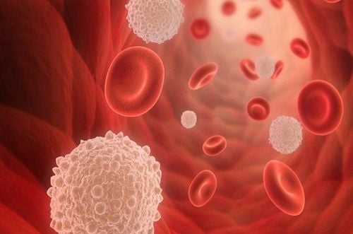
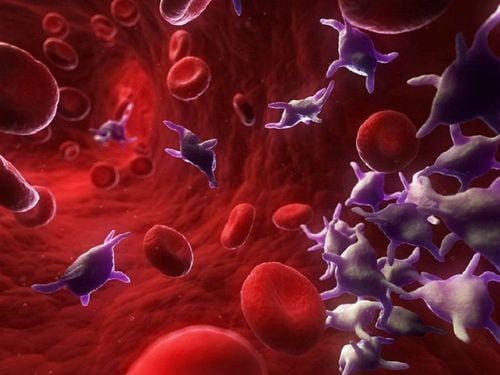
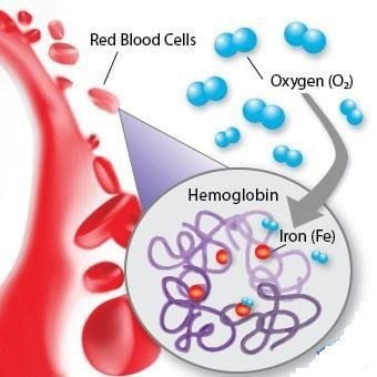
Red blood cells are cells responsible for transporting oxygen in the body. They are very small and shaped like donuts. The average lifespan of red blood cells is 120 days in the body. Red blood cells contain a protein called hemoglobin, which contains iron, which has the ability to attach oxygen and is also what gives blood its red color. Lack of hemoglobin levels is the cause of anemia.2. How are red blood cells born?
Red blood cells are born from hematopoietic stem cells, proliferate and differentiate through stages. From erythrocyte progenitor cells (CFU-E), under the influence of erythropoietin (EPO), progenitor cells of red blood cells are produced, called proerythroblasts. It is a large cell (about 20-25μm in diameter) with a round nucleus, a bright and very fine nuclear grid with a pale nucleus, sometimes the nucleus is not clearly visible. The cytoplasm is round, strongly basophilic (dark blue) because it contains many ribosomes necessary for the synthesis of large amounts of hemoglobin. A perinuclear light space is often seen on Giemsa staining slides. Sometimes, the cytoplasm is seen to protrude to form a "pseudocyst".
One pro-erythroblast produces two basophils (erythroblasts) I and four basophils II. However, under the light microscope, it is not possible to distinguish between basophilic erythrocytes I and basophils II. Its more mature (differentiated) property than pro-erythrocytosis is demonstrated by the special condensation of the artificial substance into small dark clusters, very evenly arranged in a "wheel spokes" shape Cell size cells are smaller than pro-erythroblasts (16-18 μm in diameter). Hemoglobin synthesis has only just begun at a very low rate, so on Giemsa staining slides almost no change in cytoplasmic color can be seen.
One basophilic erythrocyte gives rise to two erythroblast polycromatophils. Because the cytoplasm has already had a significant amount of hemoglobin (orange) synthesized and ribosomes still exist (basic), the cytoplasm has a mixed color of blue and orange (gray-green) on the specimen. Giemsa staining. Cells are smaller, about 12-15 μm in diameter. The nucleus is usually round, located in the center of the cytoplasm, and the nuclear chromophore begins to coagulate, creating the appearance of regular "clumps". Normally, polychromic erythrocytes have a very sharp perinuclear luminosity. This is the last stage of cell replication in the process of red blood cell differentiation. Erythroblast acidophilic erythrocytes are produced by duplication of polymorphonuclear erythrocytes. At this stage, the synthesis of hemoglobin is almost complete, the cell no longer divides. Eosinophilic erythrocytes are 10-15 μm in diameter. The nucleus is round, small, very dark in color, located in the center of the cell, and is about to dissolve. The cytoplasm is orange, the color is similar to that of mature erythrocytes.
Reticulocytes are the final stage of erythrocyte maturation with residual nuclear traces. Cell size is equal to or slightly larger than that of mature erythrocytes (7-11 μm in diameter). Human has disappeared. A few mitochondria and ribosomes remain in the cytoplasm, allowing the cell to still synthesize some hemoglobin. When staining cells with a special precipitation method (cresyl blue staining), residual nuclear traces that are reticular or dark purple-colored granules can be observed on a pale green cytoplasmic background. Reticulocytes stay in the bone marrow for about 24 hours before being released into the peripheral blood. There, they persist for another 24-48 hours in a "retic" state and then lose their nucleus completely to become mature red blood cells.Reticulocytes in peripheral blood are considered to be present in the presence of erythrocyte production. When the reticulocytes increase, the bone marrow is making strong red blood cells
In peripheral blood, the normal mature red blood cell count in healthy people is in the range of 4.0-6.0 X 10¹²/ l, corresponding to hemoglobin 120 - 180 g/l Mature red blood cells are biconcave disc-shaped, 7-8 μm in diameter, 1-3 μm thick, without nucleus. On Giemsa-stained blood smear, red red-pink catch bridge, there is a circular light in the middle.
3. What is the role of red blood cells?
What is the role of red blood cells? This is a question that many readers are interested in. The function of red blood cells is to transport oxygen (O2) from the lungs to the tissues, go everywhere in the body, and receive carbon dioxide (CO2) from the tissues back to the lungs for elimination. Red blood cells are very flexible. The role of red blood cells is to penetrate even the smallest blood vessels (capillaries), to provide oxygen to all cells in the body. When old red blood cells are destroyed in the spleen, the bone marrow will make new red blood cells to replace and maintain the lost red blood cells. When the body lacks red blood cells, it will show fatigue, blue skin due to lack of oxygen.
A blood test for red blood cells should be done when there is a lack of red blood cells. At the same time, you should have a general health examination to help detect the deficiency of red blood cells early to have a timely adjustment plan.
Please dial HOTLINE for more information or register for an appointment HERE. Download MyVinmec app to make appointments faster and to manage your bookings easily.


