This is an automatically translated article.
The article is professionally consulted by Master, Doctor Nguyen Thi Ngoc - General Internal Medicine - Endocrinology - Department of Examination & Internal Medicine - Vinmec Central Park International General Hospital.
Neuroendocrine tumors can appear in many different locations in the body, so the diagnosis is relatively complicated and can take a certain amount of time. Treatment for neuroendocrine tumors will be selected appropriately after the diagnostic process.
1. Diagnosis of neuroendocrine tumors
To diagnose neuroendocrine tumors, the doctor will need to take the history and symptoms of the disease, perform a physical examination, and order different laboratory tests and techniques, based on the specific disease case.Take history, clinical examination Take history and disease manifestations: the doctor will take the patient's medical history and disease manifestations, as well as family history. Patients should provide their doctor with full information about disease manifestations, risk factors, and previous health problems, especially information about multiple endocrine neoplasia type 1 (multiple endocrine neoplasia). endocrine neoplasia type 1 - MEN 1) (if present). Family history factors The doctor will dig up information about whether there is a family member with neuroendocrine tumors or other types of cancer. Physical examination: the doctor will check blood pressure, physical examination of organs. Laboratory tests and techniques Blood count: can help detect anemia due to chronic bleeding. Blood Biochemistry: During the diagnosis of neuroendocrine tumors, blood biochemistry results will help evaluate electrolyte status as well as blood glucose levels (abnormal blood glucose levels may suggest diabetes mellitus). , problems with the pancreas, Cushing's syndrome,...) Biochemical markers: biochemical markers are different substances, including proteins and hormones, that are secreted by cells . If levels of biochemical markers are elevated in the blood or urine, it may indicate an existing neuroendocrine tumor, or that an endocrine syndrome is occurring, such as carcinoid syndrome. Biochemical markers are also used to monitor response to treatment and to monitor recurrence. Chromogranin A (CgA): is a protein found in neuroendocrine tissues and circulating in the blood. Elevated CgA levels may indicate an existing neuroendocrine tumor, since most neuroendocrine tumors have elevated CgA.
5 - HIAA (5 - hydroxyindoleacetic acid): 5 - HIAA is a substance made up of serotonin and is measured in urine. High levels of 5-HIAA may reflect symptoms that are present as a result of carcinoid syndrome or an acute carcinoid attack. However, it is also important to note that 5-HIAA can be changed by many different foods and drugs, and some types of neuroendocrine tumors do not secrete 5-HIAA.
Based on specific symptoms, other biochemical markers that may need to be checked are: calcitonin, cortisol, adrenocorticotropic hormone (ACTH), gastrin, glucagon, insulin, somatostatin, vasoactive intestinal polypeptide (VIP), pancreatic polypeptide (PP), metanephrines.
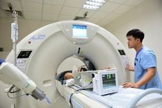
Ultrasound: Ultrasound can help detect tumors and check the functioning of the heart. Computed tomography (CT), magnetic resonance imaging (MRI): computed tomography and magnetic resonance imaging help detect the existence of tumors or not, tumors in the location. whether it has metastasized or not and with such a tumor is there any possibility of surgery. Octreotide scan: This technique uses a drug called octreotide that is similar in structure and action to a substance found in the body called somatostatin. Somatostatin binds to receptors on the surface of many neuroendocrine tumor cells, thus octreotide imaging can indicate the location of neuroendocrine tumors and indicate whether or not they have metastasized. MIBG: MIBG is an imaging technique in which metaiodobenzylguanidine (MIBG) will be introduced into the body, and then performed to determine the existence and location of some neuroendocrine tumors. certain, such as pheochromocytoma. Positron emission tomography (PET): tomography is also used to identify tumors and metastases, but is indicated when the results of other imaging techniques are unclear. . Biopsy: A biopsy means taking a sample from the tumor and taking it to pathology to check for malignancy. Biopsy is the gold standard for cancer diagnosis, as well as for tumor grading. Other subclinical techniques may be indicated: gastrointestinal endoscopy, bronchoscopy, bone scan,...

U thần kinh nội tiết nên điều trị như thế nào?
2. Treatment of neuroendocrine tumors
Treatment for neuroendocrine tumors will depend on the type of tumor, its location, and the problems caused by the overproduction of hormones.Usually the possible treatments are:
Surgery: surgery is used to remove the tumor, preferably completely removing the tumor, but in many cases only one can be removed. tumor part. Chemotherapy: Chemotherapy is used to kill tumor cells and prevent tumor recurrence. If surgery cannot completely remove the tumor, chemotherapy is a preferred option. Targeted therapy: often combined with chemotherapy to treat complex neuroendocrine tumors. Peptide receptor radionuclide therapy (PRRT): This is a special method that uses a drug containing a radioactive drug to target cancer cells, so that it can be directed directly to cancer cells. destroy cancer cells in situ with radiation (this is the advantage of PRRT compared to traditional external radiation therapy, which is both optimal and safe for healthy tissues). The drug for PRRT is lutetium Lu 177 (Lutathera) A drug that controls excessive secretion of hormones. Radiation therapy: Radiation therapy can kill tumors, but not all neuroendocrine tumors respond to radiation. Radiation therapy is an option that can be used if surgery is not feasible. Other treatments may be indicated on a case-by-case basis. Master, Doctor Nguyen Thi Ngoc has more than 10 years of studying, researching and working in the field of endocrinology. Doctor Ngoc graduated with a Master's degree in General Internal Medicine from Hanoi Medical University and studied as a Resident Doctor at Lyon University (France). Currently, Dr. Ngoc is a treating doctor at the Department of Examination and Internal Medicine, Vinmec Central Park International General Hospital.
Please dial HOTLINE for more information or register for an appointment HERE. Download MyVinmec app to make appointments faster and to manage your bookings easily.
Articles refer to sources: mayoclinic.org and webmd.com
MORE:
When to have a full body magnetic resonance imaging (MRI) scan? How many radiation treatments are there? Which option is reasonable? What is cancer? The difference between cancer cells and normal cells