Patient T (female, 56 years old), after self-detecting an abnormal mass in the right breast, visited another medical facility. Through examination and core needle biopsy, the patient was diagnosed with invasive carcinoma – non-specific type, grade III, with a tumor size of 15x35mm. However, just 3 days after the biopsy, when the breast tissue was still swollen and bruised, the patient was prescribed a breast MRI.
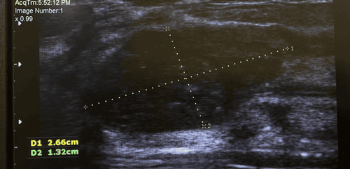
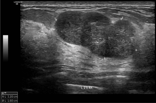
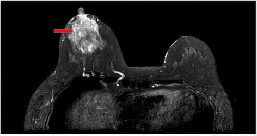
Subsequently, the patient decided to visit the Breast Center at Vinmec Times City Hospital for consultation and in-depth examination. Here, the doctors re-evaluated the MRI film and found that the actual tumor size was larger than the previous diagnosis, approximately 45x33x35 mm, extending close to the nipple and showing signs of hemorrhage and edema. This limited the accurate assessment of the disease stage and invasion level, affecting the patient's ability to preserve the nipple.

To clarify the extent of the damage, doctors ordered immunohistochemical staining on the biopsy sample and performed a right axillary lymph node biopsy to assess the spread of cancer cells. Through thorough examination and consultation, the patient was definitively diagnosed with stage IIB breast cancer, Her2-positive (Her2+), a type of breast cancer characterized by the presence of the Her2 protein on the cell surface, which makes cancer cells grow and spread faster.
After a comprehensive evaluation of the patient's condition, the case was discussed at the Vinmec Times City Multidisciplinary Oncology Council, and a neoadjuvant treatment plan (chemotherapy before surgery) was agreed upon according to the TCHP regimen. This regimen combines chemotherapy and antibodies to shrink the tumor size, reduce invasion, and create the most favorable conditions for radical surgery later.
Breast cancer diagnosis requires a combination of many factors, from equipment and machinery to the expertise of doctors. The above case is a typical example showing the important role of breast MRI in diagnosing breast cancer, especially before performing a biopsy. If the patient had continued to wait without re-examination at the Breast Center, Vinmec Times City Hospital, she would have faced the risk of not being able to preserve the nipple and encountering more difficulties in treatment.
By using MRI, the images of the lesion will more accurately reflect the structure and characteristics, minimizing interference from post-biopsy swelling or bleeding. Additionally, performing MRI at least 7–10 days after the biopsy helps the images to be clearer and greatly supports effective treatment planning. The Vinmec Times City Breast Center is one of the medical units equipped with full facilities for cancer diagnosis and screening, especially breast cancer, such as 2D, 3D ultrasound machines, 2D, 3D mammography, breast MRI machines, and gathers leading oncology experts capable of optimizing diagnostic and treatment results for patients.