This is an automatically translated article.
Posted by Doctor Vu Ngoc Thang - Department of General Surgery - Vinmec Times City International Hospital
Prostate cancer is a disease only seen in men, the prostate gland is located below the bladder, in front of the large intestine. This is a slow-growing disease, most people with mild prostate cancer can live for many years if detected early. After all, if the disease is severe, it will spread very quickly and can cause death.
Here are the methods to diagnose prostate cancer :
1. Finger rectal examination
Digital rectal examination is the method used to detect disease. Whitmore 1956, was the first to use the concept of "palpable fingertip" for prostate cancer. Jewett 1956, published the rate of prostate cancer 50% at prostate biopsy for suspicious cases during rectal examination, nearly 10 years later the author was the first to classify prostate cancer stage based on rectal examination.
Rectal examination is a simple, quick, and low-cost examination method, but there are many reasons that make the diagnostic sensitivity not high.
2. P.S.A
PSA is a prostate-specific antigen, but PSA exists in a number of other glandular organs, so it is still not an ideal specificity for prostate cancer. The higher the PSA result, the more suspicious it is for prostate cancer. Currently, PSA is still considered the most effective in diagnosing, staging and monitoring the development of prostate cancer, especially newly published studies show that PSA can identify high-risk cases. chance to develop into prostate cancer in the future.
The advantage of PSA is that it is objective, the results do not depend on the examiner, avoiding the patient's embarrassment and fear of pain. In many studies, the authors also confirmed that the use of PSA increases the chance of early detection of the disease, which is potentially curable. PSA is a good tool to predict risk, monitor and evaluate outcomes after treatment.
The new role of PSA today as a determining factor in the risk of prostate cancer in the future. PSA can predict Cancer 25 - 30 years in advance.

Kết quả PSA càng cao càng nghi ngờ ung thư tuyến tiền liệt
3. Ultrasound
Ultrasound is a valuable diagnostic measure for prostate cancer, ultrasound is an important support for clinical examination, so it is necessary to have people who specialize in urological ultrasound make the diagnosis. In developed countries, when studying screening and diagnosis in depth, it has been shown that transrectal ultrasound has the ability to detect prostate cancer tumors with a volume of 2 - 4mm.
Ultrasound of the pubic bone :
Ultrasound of the pubic bone allows to evaluate the effects of prostate cancer on the upper urinary tract, especially in the late stages of the disease. The bladder wall can be dilated thin, or inflamed, the ureter of the renal pelvis dilated with water retention due to compression of the tumor. Through ultrasound, other lesions such as pelvic lymph nodes can be assessed, the extent of tumor invasion into the bladder, and prostate size can also be measured. The disadvantage of ultrasound on the pubic bone is that it uses a low-frequency transducer, when the ultrasound is limited to the pubic bone, it cannot directly observe the prostate gland, so it does not give clear images.
Transrectal ultrasound:
Since 1968, Watanabe has used transrectal ultrasound for the first time to describe the image of the prostate gland. This technique has been widely used in clinical practice since the mid-1980s. Transrectal ultrasound uses a high-frequency probe of 5-7MHz, so it gives a clearer image than the suprapubic ultrasound, which can detect can now detect UT 2-4 mm in diameter, and make biopsies more accurate thanks to the locator that comes with the probe.
4. Biopsy of the prostate
Indications for prostate biopsy are guided by rectal examination and blood PSA levels.
Biopsy can be done in several ways:
Transrectal biopsy under finger guidance or transrectal ultrasound. Perineal biopsy with or without ultrasound guidance. Transrectal prostate biopsy under ultrasound guidance is the technique of choice today. Usually, each lobe of the gland is taken from 3 to 6 pieces from the top to the base of the gland, numbering the positions to be able to map the pathological lesions of prostate cancer.
The combination of rectal examination, periodic PSA blood testing, and prostate biopsies when in doubt allows the diagnosis of >90% of adenocarcinomas at localized stage and contributes significantly to the effectiveness of treatment. treat.
5. Some diagnostic imaging methods
Bone scintigraphy, magnetic resonance imaging, PET Scan, computed tomography, ultrasound, bone X-ray, intravenous drug nephrography, are considered methods to determine the development stage of prostate cancer.
5.1. Computed tomography (CT scan) or Magnetic Resonance (MRI)
- CT scanner : with the continuous development of imaging diagnostics, spiral CT was born, since 1990 with 1 head, rotates 3600, takes snapshots, clear 3-D images, has been further improved up to now. 64 to 320 heads, significantly increase the number of images taken in 1 second, making it an ideal imaging method in determining tumor invasion, lymph node metastasis, and vascular distribution to help guide direction. To solve.
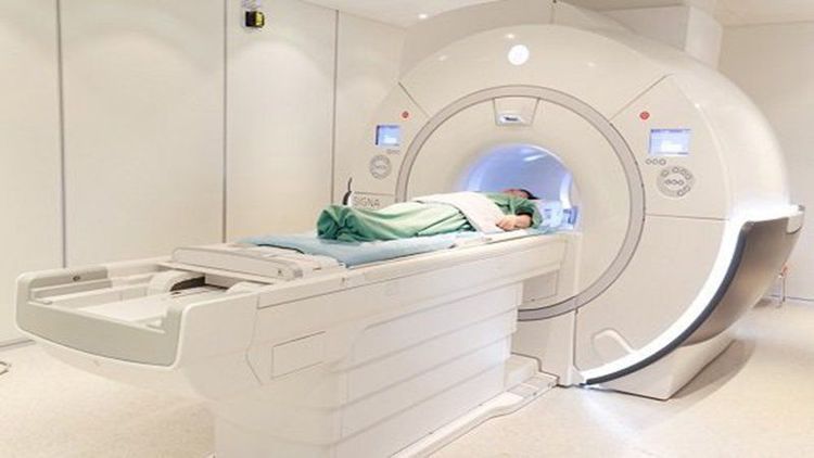
- MRI: Very valuable in diagnosing the extent of tumor invasion into surrounding tissues and regional lymph nodes, especially intrarectal MRI. Diagnostic value of intrarectal MRI for prostate cancer: sensitivity 53%, specificity 94%, accuracy 70%, and helps prostate biopsies be more accurate.

Chụp MRI giúp chẩn đoán ung thư tuyến tiền liệt
5.2. Scintigraphy osseuse
Bone scintigraphy has high sensitivity in the diagnosis of bone metastases. Bone scan should be combined with bone examination for differential diagnosis with other bone pain symptoms such as osteoarthritis, disc herniation... Positive bone scan is not specific for prostate cancer, scan results Negative bone is very valuable in the assessment before and after treatment.
5.3. PET Scan (positron emission tomography)
PET Scan is a modern method of staging the disease based on tomography images taken after injecting radioactive substances such as 18 - Fluoro - Deoxy Glucose (FDG). These radioactive substances will accumulate in the sites of increased glycolysis due to UT. The advantage of this method is the ability to detect metastases early.
Vinmec Times City International General Hospital now has an Intensive Prostate Cancer Care Package to help customers detect the possibility of prostate cancer early, helping customers take preventive measures; Detect the disease at an early stage, without typical symptoms to receive early treatment and prevent complications.
Prostate cancer intensive examination package for customers:
Men aged 50 years or older, PSA index >4 or/and suspected by perianal prostate examination Having symptoms following: Urinary tract obstruction: dysuria, dysuria, hematuria, sometimes urinary retention; Pelvic discomfort, penile pain (rare). Customers wishing to be examined at Vinmec International General Hospital can register HERE.
References:
American Society of Radiology : PIRADS score v2.1 full version (2019) : https://www.acr.org/-/media/ACR/Files/RADS/Pi-RADS/ PIRADS-V2-1.pdf?la=en NCCN Guidelines, Prostate Cancer, Version I.2018.


