This is an automatically translated article.
The article is made by Master, Doctor Ton Nu Tra My - Department of Diagnostic Imaging - Vinmec Central Park International General Hospital.
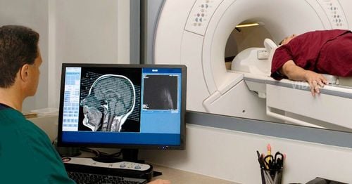
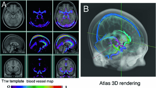
Magnetic resonance angiography of the intracranial vasculature with injection of magnetic contrast agent is a type of cranial magnetic resonance focusing on intracranial blood vessels and using magnetic contrast agent, giving very clear images of the brain. Vascular and cerebral blood flow help to accurately assess cases of suspected neurovascular disease.
1. What is an intracranial magnetic resonance angiogram with injection of magnetic contrast?
Magnetic resonance imaging of the intracranial vasculature with injection of magnetic contrast is a technique that uses magnetic fields and pulses of radio wave energy and uses intravenous contrast agents for the purpose of surveying the carotid system - effective intracranial background. This method allows to examine both the arterial and venous systems of the brain, capable of detecting many cerebrovascular diseases such as stenosis, arteriovenous malformations, aneurysms, cerebral venous malformations or blood vessels. block arteries as well as veins.
This technique is an effective technique to examine the vertebral and intracranial carotid system, with higher accuracy than examining the cerebrovascular system by magnetic resonance without injecting contrast. However, adverse events associated with contrast agents may occur.
2. In which cases is an intracranial magnetic resonance angiogram with injection of magnetic contrast agent indicated?
The intracranial angiogram is not clear enough on the non-injection film; The doctor suspects that the patient has intracranial vascular diseases such as:: cerebrovascular (arterial or venous) malformations, aneurysms, intracranial and extracranial cerebral stenosis; Cerebral stroke: Infarction to find the location of large vessel occlusion, cerebral hemorrhage but need to find aneurysm, vascular malformation, carotid artery - cavernous sinus...; Look for intracranial venous sinusitis; Evaluating the effectiveness of post-treatment in cases of cerebral aneurysm node, cerebral arteriovenous malformation...

3. People who are not allowed to have magnetic resonance imaging
Absolutely contraindicated for patients with pacemakers in the body
Consider taking pictures in some of the following cases:
In people with magnetic metal; People with liver and kidney failure, pregnant and lactating women should consider injecting contrast agents; Inform the doctor about the history of food allergy, asthma, etc. because these subjects are at risk of hypersensitivity, anaphylaxis to contrast; People who are afraid of light, afraid of lying alone, cannot lie still.
4. Magnetic resonance imaging procedure of intracranial vascular system with injection of magnetic contrast agent
4.1 Preparation
Drugs: tranquilizers, contrast agents, anti-shock boxes; Patients do not need to fast, hear clearly explained from the doctor about the purpose and possible complications; The patient needs a written request for magnetic resonance imaging, a written commitment to consent to the injection of contrast; Instruct the patient to change clothes of the MRI room and remove contraindicated items such as watches, jewelry, metal, and cell phones. 4.2 Procedures
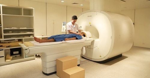
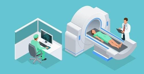
Step 1: Move the patient into the machine compartment. The patient is placed supine on the imaging table and asked to remain still. People who are not able to lie still like children, stimulants need to be sedated to ensure they stay still during the scan. Then move the table into the engine compartment.
Step 2: The technician performs the technique from the remote control room. Techniques include:
Location capture; Select diagnostic pulse sequences suitable for examination purposes. Perform common pulse sequences: T1, T2, Flair for all objects with multiple cutting directions including horizontal, horizontal and vertical; Selection of pulse sequences for the suspected pathology to be searched. For example, T2* pulses for bleeding lesions, diffusion pulses for ischemic-related lesions; Non-injectable pulse train (TOF3D) can also be performed.
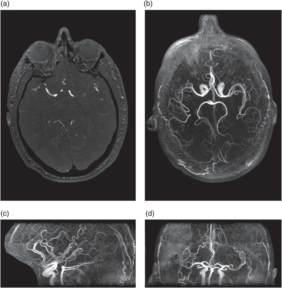
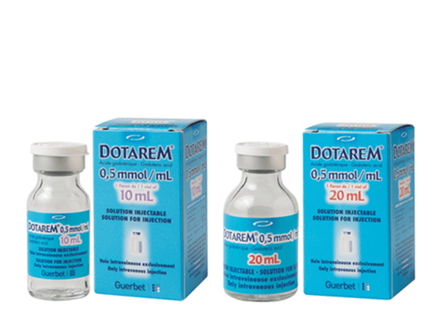
Step 3: Inject contrast drug. DSA-MRI angiogram: Select the pulse sequence of the DSA pulse, get the positioning frame for the head region. The drug is then injected through the electric syringe and pulsed.
Step 4: Process the image, read the lesion.
5. Complications of technology
Nephronic fibrosis is a rare complication associated with the administration of gadolinium contrast media. It usually occurs in patients with severe kidney disease. Therefore, careful assessment of renal function is required before considering contrast injection. There is a very slight risk of allergic reactions if contrast materials are used. Such reactions are usually mild and controlled with medication. The most severe condition is anaphylaxis, which can cause rapid blood vessel destruction and death. It is necessary to ask carefully for an allergy history, have adequate anti-shock equipment and have a timely management attitude. Strong magnetic field is not harmful. However, it can cause implantable medical devices such as pacemakers to malfunction or affect film image quality. Vinmec International General Hospital is currently one of the major hospitals with modern machinery and equipment for general medical examination and treatment and accurate and modern MRI for brain and vascular diseases. brain in particular.
Especially, Vinmec International General Hospital is the first unit in Southeast Asia to put into use the new 3.0 Tesla Silent Resonance Imaging machine from the US manufacturer GE Healthcare.
The machine currently applies the safest and most accurate magnetic resonance imaging technology available today, without using X-rays, non-invasive. Silent technology is very beneficial for patients who are young children, the elderly, patients with weak health or have just had surgery.
Master. Dr. Ton Nu Tra My is currently a Doctor of Radiology, Vinmec Central Park International General Hospital. Dr. Tra My used to be a lecturer in the Department of Diagnostic Imaging, Hue University of Medicine and Pharmacy and had a long time working at the Department of Diagnostic Imaging, Hue University of Medicine and Pharmacy Hospital.
Please dial HOTLINE for more information or register for an appointment HERE. Download MyVinmec app to make appointments faster and to manage your bookings easily.
Reference source: radiologyinfo.org


