This is an automatically translated article.
The article is made by Master, Doctor Ton Nu Tra My - Department of Diagnostic Imaging - Vinmec Central Park International General Hospital.
Magnetic resonance angiography without injection of magnetic contrast is a technique that uses specific pulse sequences of magnetic resonance in the evaluation of intracranial vessels without the need for injection of magnetic contrast.
Magnetic resonance angiography allows images of blood vessels and blood flow to be of the same quality as conventional angiography, but is not an invasive procedure to the blood vessels to evaluate narrowing, cerebrovascular obstruction, cerebral aneurysm as well as other pathologies.
1. Learn about magnetic resonance imaging of the intracranial vasculature without injection of magnetic contrast agent
Magnetic resonance angiography of the intracranial vasculature without injection of magnetic contrast is a technique that uses magnetic fields and pulses of radio wave energy, without the use of magnetic contrast agents to provide clear images of cerebral blood vessels. At best, conventional cranial magnetic resonance imaging cannot provide a good picture of blood vessels and blood flow to help identify abnormalities.
This technique is widely applied in neuropathies with suspected abnormalities of blood vessels as well as circulation, not affected by radiation, for the purpose of investigating the carotid system - effective intracranial background. fruit.
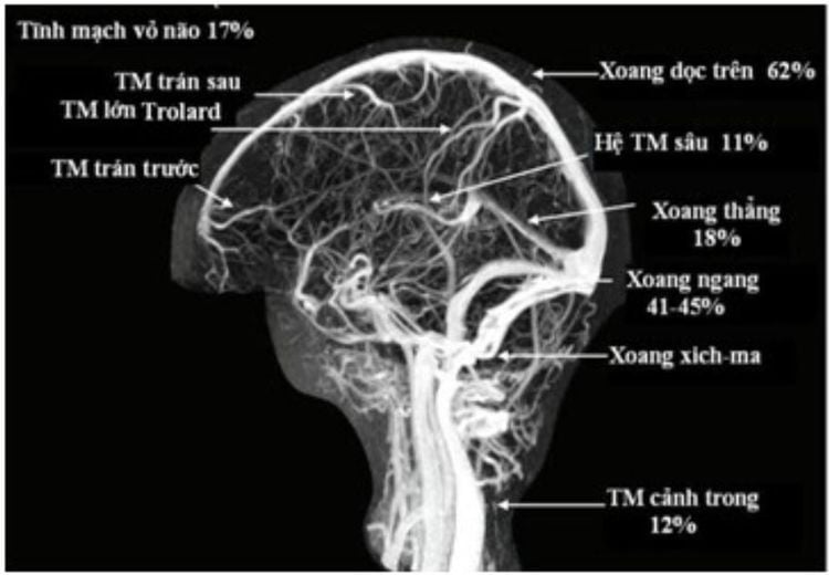
This method allows to examine both the arterial and venous systems of the brain, capable of detecting many cerebrovascular diseases such as stenosis, arteriovenous malformations, aneurysms, cerebral venous malformations or other diseases. arterial as well as venous thrombosis.
2. Advantages of vascular magnetic resonance imaging without injection of magnetic contrast agent
As a non-invasive imaging technique, there is no radiation exposure. The technique gives detailed images of the morphology and flow signals of the major blood vessels of the brain. Magnetic resonance angiography without contrast is less time-consuming and less expensive than angiography and requires no recovery time. Without the use of contrast, it can still provide clear images of blood vessels. This is very valuable in patients who are allergic to contrast agents or patients with impaired liver or kidney function.
3. Indications and contraindications of intracranial magnetic resonance angiography without injection of magnetic contrast
3.1 Indications Suspected cerebral aneurysm or arteriovenous malformation (AVM), venous malformation; Suspected cerebral stenosis, intracranial and extracranial occlusion; Stroke: Cerebral infarction to find the location of large occlusion, brain bleeding to find aneurysms, vascular malformations, carotid artery - cavernous sinus fistula indirectly, directly...; Finding intracranial venous sinusitis...; Screen for arterial disease, especially in patients with a family history of the disease. Assess the pulse after treatment such as post-nodal examination of cerebral arteriovenous malformations, cerebral aneurysms, and disease progression (eg, checking venous sinus recanalization due to thrombosis after treatment...). 3.2. Contraindications Patients with pacemakers (absolute contraindications); In humans with ferromagnetic metals (relative contraindications); People are afraid of light, afraid of lying alone, unable to lie still.

4. What should the patient pay attention to?
Women who are pregnant need to inform their doctor, although there are no studies that indicate an adverse effect of magnetic resonance on pregnant women or the fetus. However, when scanning the fetus will be in a strong magnetic field, so pregnant women in the first 3 months should not have an MRI. For infants and young children who are unable to lie still during the scan, sedation or anesthesia is required. People with photophobia, anxiety, and agitation may also ask their doctor to use a sedative before the scan. Remove jewelry, accessories, electronic metal, and cell phones as these can be affected by magnetic fields. If the patient has a pacemaker on the body, do not undergo magnetic resonance imaging.
5. Magnetic resonance imaging procedure of intracranial vascular system without injection of magnetic contrast agent
5.1 Working principle of magnetic resonance Magnetic fields are created by passing an electric current through a coil. The coils are placed in the machine for the purpose of sending and receiving radio waves. Radio waves rearrange hydrogen atoms that exist naturally in the body. When the hydrogen atoms return to their normal alignment, they emit different amounts of energy depending on the type of body tissue. The scanner captures this energy and creates an image using this information, each showing a thin slice of the body.
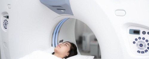
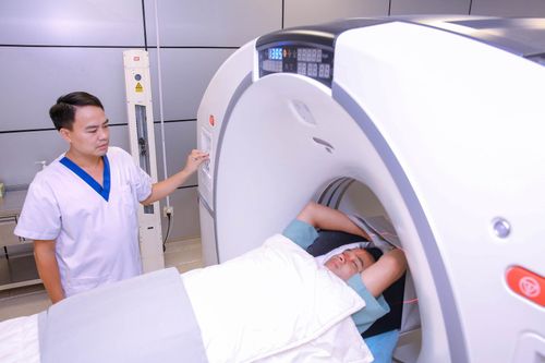
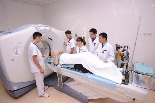
5.2 Preparation Instruct the patient to change into the dressing room and remove items. Have a clinician's order for a scan with a clear diagnosis. 5.3 Procedure Step 1: Move the patient into the machine compartment. The patient is placed supine on a movable examination table. Straps and washers can be used to help you stand still and maintain the patient's position and then move the patient into the machine compartment.
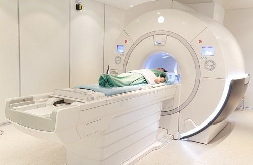
Step 2: The technician will take pictures at the remote control room
Location capture; Selection of diagnostic pulse sequences suitable for examination purposes; Do the usual pulse sequences: T1, T2, Flair for all subjects. The cutting direction includes horizontal, horizontal and vertical cutting; Selection of specific pulse sequences for the particular pathology to be sought. For example, T2* pulses for bleeding lesions, Diffusion pulses for brain infarction-related lesions...; 3D TOF pulse imaging for the arterial system and 3D PC pulse for the venous system. Step 3. Process the images obtained on the workstation screen, select the necessary images to reveal the pathology for film printing.
Step 4: Read the results and conclusions from the radiologist.
Vinmec International General Hospital is currently one of the major hospitals with modern machinery and equipment for general medical examination and treatment procedures and accurate and modern MRI for brain and vascular diseases. brain blood in particular.
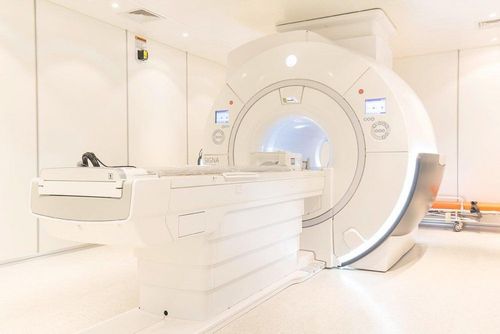
Especially, Vinmec International General Hospital is the first unit in Southeast Asia to put into use the new 3.0 Tesla Silent Resonance Imaging machine from the US manufacturer GE Healthcare.
The machine currently applies the safest and most accurate magnetic resonance imaging technology available today, without using X-rays, non-invasive. Silent technology is very beneficial for patients who are young children, the elderly, patients with weak health or have just had surgery.
Master. Dr. Ton Nu Tra My is currently a Doctor of Radiology, Vinmec Central Park International General Hospital. Dr. Tra My used to be a lecturer in the Department of Diagnostic Imaging, Hue University of Medicine and Pharmacy and had a long time working at the Department of Diagnostic Imaging, Hue University of Medicine and Pharmacy Hospital.
Please dial HOTLINE for more information or register for an appointment HERE. Download MyVinmec app to make appointments faster and to manage your bookings easily.
References: strokecenter.org, radiologyinfo.org


