This is an automatically translated article.
The article is professionally consulted by Master, Doctor Nguyen Viet Thu - Doctor of Radiology and Nuclear Medicine - Department of Diagnostic Imaging and Nuclear Medicine - Vinmec Times City International General Hospital. Dr. Nguyen Viet Thu has more than 20 years of experience working in the field of diagnostic imaging, former Secretary of the Hanoi Department of Radiology, directing the lower level in the field of Diagnostic Imaging.Lung cancer is a dangerous disease with a high mortality rate. However, if the disease is diagnosed and treated early, the prognosis is very good, reducing the risk of death. Currently, there are many methods to diagnose lung cancer such as computed tomography (CT scanner), magnetic resonance imaging (MRI), PET/CT scan, biopsy, bronchoscopy, endobronchial ultrasound manage. Treatment options depend on the extent of the disease, and include surgery, radiation therapy, targeted therapy, and systemic therapy or a combination of methods.
1. Lung cancer and what you need to know
Lung cancer forms in the tissues of the lungs, usually in the cells that line the airways. About 85% of lung cancer tumors occur in current or former smokers. In addition, other risk factors for lung cancer may include: Age, exposure to secondhand smoke, exposure to asbestos or radon gas, genetic factors.Typical symptoms of lung cancer depend on the location and extent of the cancer, but it can also include shortness of breath, chest pain, chronic cough, hemoptysis, shoulder pain chronic, hoarse voice, difficulty swallowing, unexplained weight loss, fatigue, unusual bone pain. In most cases, early lung cancer has no symptoms and it can only be detected based on the results of imaging tests. If lung cancer spreads to the brain, patients may experience blurred vision, seizures, headaches, or symptoms of a stroke.
There are two main types of lung cancer with different microscopic manifestations: Small Cell Lung Cancer (SCLC) is usually found in active or former smokers. Although SCLC is a less common type of lung cancer, it is highly malignant and is more likely to metastasize to other sites in the body. Non-small cell lung cancer (NSCLC) tends to grow more slowly and metastasize outside the lungs longer.

2. Measures to diagnose lung cancer
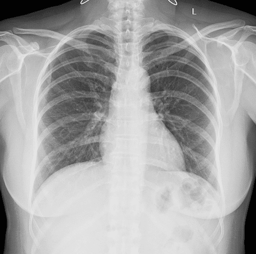
A diagnosis of lung cancer begins with understanding your medical history, risk factors, symptoms, and evaluation tests. Even if there are no symptoms, patients should have regular lung cancer screening tests. The most common lung cancer screening and diagnostic tests are the following imaging tests:Low dose computed tomography (LDCT): This is a combination of low-dose CT scans that produce low-dose computed tomography (LDCT). images of sufficient quality to detect many lung diseases and detect other abnormalities. The amount of ionizing radiation in a low-dose computed tomography scan will be up to 90% less than the amount of ionizing radiation in a standard chest CT scan. Cardiopulmonary X-ray: With a simpler means than CT, it is commonly used to examine the thorax, heart and mediastinum. This is a low-dose X-ray test and is also the first type of imaging test done when a patient has respiratory symptoms. On the chest X-ray film, the doctor can detect tumors, nodules, abnormal opacities in the lungs, evaluate the heart's enlargement, or pleural effusions... is a non-invasive technique. provides a baseline image of the thorax. If a tumor is suspected or an undetermined lesion is present, the patient will be advised to combine it with a chest CT scan. Sputum cytology: This diagnostic test is done by taking a sample of sputum (coughed up mucus) and examining it under a microscope to determine if abnormal cells are present. Testing for cancer cells in a sputum sample can be useful in detecting lung cancer early. But this is a test that provides information about the cancer cells on the slide, so it needs to be done with good technical standards.
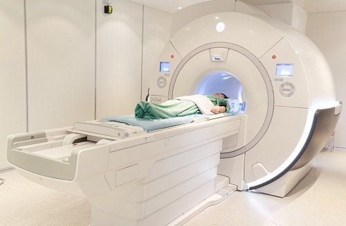
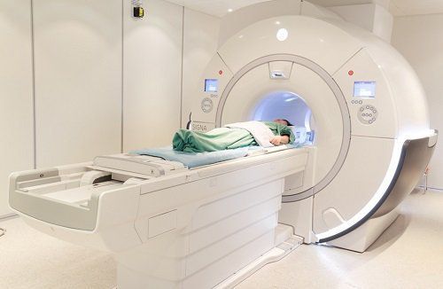
Chest CT: with conventional dose, or high resolution with or without intravenous contrast, may be performed to view The finer details inside the lungs and detecting tumors can be difficult to see on a conventional X-ray. It is also used for a more detailed evaluation of underappreciated abnormalities on low-dose CT. Chest CT is the primary imaging test to evaluate for lung cancer. CT scans of the abdomen and pelvis may also be performed when evaluation for secondary lesions is needed. PET/CT tomography: This is a test that uses both a PET machine in combination with a CT machine and a small amount of radioactive drug (Fluorodeoxyglucose, FDG), to help pinpoint the exact location and extent of the cancer. lungs, and assist in the assessment of outcomes after treatment. PET/CT scan is also effective in detecting metastatic lesions in bones, pleura, lymph nodes and lesions in other organs. Bronchoscopy: This test is indicated for most cases of lung tumors. The process of bronchoscopy is to visually examine the inside of the airways (trachea and bronchial tree) using a rigid or flexible tube with a light that is inserted into the windpipe through the nose. Endobronchial ultrasound: Using a bronchoscope probe, determines tumor invasion and guides biopsy, in addition, also evaluates adjacent lymph nodes in the field of view of the probe. Cranial MRI: performed to evaluate tumors that have spread to the brain. Chest MRI: Chest MRI is uncommon in lung cancer. But it can give us detailed images of the mediastinum, chest wall, pleura, heart, and blood vessels. As a result, doctors may be able to evaluate features of the tumor, including cancer of the lung or other tissues, that are not fully evaluated by other imaging modalities (such as CT). Chest MRI is also useful for patients with contraindications to standard imaging tests.
After having an accurate diagnosis of the disease and stage, the patient will be consulted to choose the most appropriate and effective treatment methods. The treatment process is always closely coordinated with many specialties: Diagnostic Imaging, Biochemistry, Immunology, Cardiology, Stem Cell and Gene Technology, Obstetrics and Gynecology, Endocrinology, and Medicine. Rehabilitation, Psychology, Nutrition... to bring the highest efficiency and comfort to the patient. After undergoing the treatment phase, the patient will also be monitored and re-examined to determine whether the cancer treatment is effective or not.
Please dial HOTLINE for more information or register for an appointment HERE. Download MyVinmec app to make appointments faster and to manage your bookings easily.
Reference source: radiologyinfo.org


