This is an automatically translated article.
The article is professionally consulted by Master, Doctor Nguyen Van Huong - Department of Diagnostic Imaging - Vinmec International Hospital Da Nang. The doctor has many years of experience in the field of diagnostic imaging.1. Pneumonia and things to know
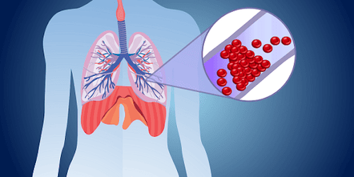
Pneumonia is an inflammatory infection caused by a virus, bacteria, fungus, or other germs. To diagnose pneumonia, your doctor will perform a physical exam and take a chest X-ray, chest CT, chest ultrasound, or lung biopsy. In addition, the doctor may further evaluate lung condition and function with thoracoscopy, placement of a chest drain, or drainage of an abscess with imaging.Patients with pneumonia may have the following signs:
Cough with phlegm or sometimes blood Fever Shortness of breath or difficulty breathing Feeling chills or shaking Fatigue Sweating Chest or muscle pain Young children, adults 65 years old and people with existing health problems are at high risk for pneumonia. Factors that increase the chance of getting pneumonia include:
People with conditions such as emphysema, HIV/AIDS or other lung diseases, conditions that affect the immune system
People with the flu People with exposure to and inhalation of various chemicals People who smoke or drink too much alcohol.

Treatments for pneumonia may include:
A thoracentesis is a way of taking fluid from a patient's chest cavity for study to help the doctor determine which germs are causing the illness. X-ray, CT, or ultrasound may be used to guide thoracoscopy. Fluids will also be removed during this procedure which will help relieve symptoms. Chest tube placement is an intervention that involves inserting a thin plastic tube into the pleural space (the area between the chest wall and the lungs to remove excess fluid or air.) The procedure is performed under CT guidance. or ultrasound Draining the abscess If an abscess has formed in the lung, it can be drained out by inserting a small drainage tube (catheter) into the abscess cavity under CT Scan or ultrasound guidance. The sound helps to put the needle directly into the abscess cavity and then insert the drain to remove all the pus in the abscess.
2. Ultrasound, CT/MRI in evaluating pneumonia
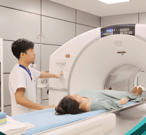
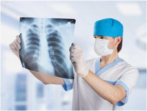
To make a diagnosis, the doctor will begin to learn about the patient's medical history and symptoms. The patient is then examined for physical symptoms. When checking for pneumonia, your doctor will listen for unusual sounds such as crackles, crackles, or wheezing. If the patient is diagnosed with possible pneumonia, the doctor will recommend imaging tests to support the diagnosis. One or more of the following tests are usually ordered to evaluate for pneumonia:Chest X-ray: An X-ray exam that will allow your doctor to see your lungs, heart, and blood vessels to help determine your condition. pneumonia. When reading the X-ray results, your doctor will look for white spots in the lungs (called infiltrates) to identify an infection. This examination will also help determine if the patient has any complications related to pneumonia such as an abscess or pleural effusion (fluid surrounding the lungs). Lung CT: A CT scan of the chest may also be done to see the finer details inside the lungs. In cases where the X-ray results can be difficult to see to detect pneumonia, the doctor will choose a CT scan of the lungs. A CT scan also shows the airways (trachea and bronchi) in great detail and can help determine if pneumonia is related to a problem in the airways. A CT scan can also show complications of pneumonia, an abscess or pleural effusion, and enlarged lymph nodes. Chest ultrasound: An ultrasound may be used if fluid around the lungs is suspected. An ultrasound examination will help determine the amount of fluid present and may assist in determining the cause of the fluid. Chest MRI: An MRI or magnetic resonance imaging is an advanced imaging technique that uses a magnetic field and radio waves to create images. Due to the advantage of being able to give clear and detailed images, it is highly appreciated in the diagnosis of lung diseases as well as diseases related to other parts such as spine, nerves, musculoskeletal, etc. ...
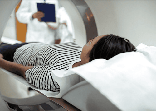
Vinmec International General Hospital is a quality medical facility in Vietnam with a team of highly qualified medical professionals, well-trained, specialized at home and abroad, and experienced. A system of modern and advanced medical equipment, possessing many of the best machines in the world such as: CT scan 460 slices, MRI 3.0Tesla helps accurately diagnose and detect many difficult and dangerous diseases in short time. Thereby supporting the diagnosis and treatment of doctors most effectively. The hospital space is designed according to 5-star hotel standards, giving patients comfort, friendliness and peace of mind.
If you have a need for consultation and examination at Vinmec Hospitals under the nationwide health system, please book an appointment on the website for service.
Please dial HOTLINE for more information or register for an appointment HERE. Download MyVinmec app to make appointments faster and to manage your bookings easily.
Reference source: radiologyinfo.org


