This is an automatically translated article.
The article was professionally consulted by Dr. Phan Dinh Thuy Tien - General Internal Medicine - Department of Medical Examination & Internal Medicine - Vinmec Nha Trang International General Hospital.Echocardiography is a non-invasive imaging technique used to evaluate and examine the patient's heart or major blood vessels. In addition, at present, Doppler echocardiography method is gradually being used in popularity because of its outstanding advantages over basic echocardiography.
1. What is Doppler echocardiography?
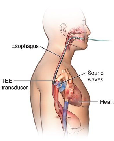
Doppler echocardiography is a transthoracic echocardiography method that helps probe the heart and large blood vessels to diagnose heart diseases, aortic diseases, congenital heart diseases,... with relatively high accuracy. .The main advantage of the method is that it is fast, it can be performed in the ultrasound department or at the bedside in emergency cases with an ultrasound probe with a frequency of 3-5 MHz for exploration. However, this method also has limitations for patients with obesity, thick chest wall, thick subcutaneous fat layer or patients with mechanical heart valves, the image quality is quite limited. Therefore, in these subjects, transthoracic ultrasound only plays a role in detecting, classifying and emergency management, then the patient is transduced into the esophagus to confirm the diagnosis and evaluate further. .
2. Who should have Doppler echocardiography?

Người bị tăng áp động mạch phổi sẽ được chỉ định siêu âm Doppler tim
Patients with lung-related diseases such as pulmonary hypertension or signs related to pulmonary hypertension. People with abnormal blood pressure, unstable increase or decrease in blood pressure suspected of cardiovascular related. History of heart disease, congenital heart defects, heart valve diseases such as valvular regurgitation, valvular stenosis require periodic cardiac function testing. Suspected tumor or thrombus abnormalities should be investigated. Unexplained cardiac arrhythmia or suspected cardiac failure. Cardiac ischemic disease or myocardial infarction leads to complications. The patient has signs of chest pain, shortness of breath and continuous and prolonged. Patients with diseases related to arteries and veins of the heart, pericardium,...
3. Key parameters in Doppler echocardiography
Through Doppler echocardiography, doctors will assess the cardiovascular status based on a number of key parameters such as:Muscle fiber shortening index (%D): is a value equivalent to EF, only assessing the basal area. of the left ventricle Ejection fraction (EF): more accurately assesses myocardial contractile function Cardiac output: assesses cardiac output and index based on flow through the aortic valve Rate of increase in ventricular pressure left ventricular maximal left ventricular function index: independent of heart rate and ventricular shape, which helps to assess total left ventricular function Maximum systolic velocity through the mitral annulus, systolic: corresponds to the displacement of the mitral annulus toward the apex of the heart during systole, reflecting the total infiltrative function of the left ventricle
4. What do Doppler echocardiogram results reflect?

Siêu âm Doppler có thể giúp các bác sĩ phát hiện tình trạng cục máu đông tại buồng tim nếu có
Abnormal signs of heart valves based on functioning status such as the ability to receive enough blood flow in diastole , valve opening or opening or closing during systole Detect tumors or blood clots in the chambers of the heart. Determine if the functions of the heart wall and the pumping state of the wall are normal. From there evaluate the damage in the heart muscle if any. Determine the pumping power of the cardiovascular system. This is an important factor to help classify the patient's heart as healthy or not. Observing the change in heart size helps to identify diseases in the cardiovascular system, abnormal abnormalities in the heart structure, problems with the heart structure. problems in the heart chambers. Raise suspicion initially when the patient has pulmonary embolism, then make a definite diagnosis when there are negative signs. Through the parameters in the Doppler echocardiogram, the doctor will assess the patient's cardiovascular health status. However, Doppler ultrasound results depend a lot on the level of the operator, the technical process and the system of machines used in the ultrasound process. Therefore, when there is a need for ultrasound, you should choose implementing prestigious and quality hospitals and medical facilities.
Vinmec International General Hospital has applied Doppler ultrasound technique in examination and diagnosis of many cardiovascular diseases. The Doppler ultrasound technique at Vinmec is performed methodically and according to the standards of the procedure by a team of highly skilled doctors, modern machinery, thus giving accurate results, greatly contributing to the quality of life. determine disease and disease stage.
If you have a need for medical examination by modern and highly effective methods at Vinmec, please register for an online examination.
Please dial HOTLINE for more information or register for an appointment HERE. Download MyVinmec app to make appointments faster and to manage your bookings easily.


