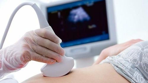
Ultrasound is an important imaging method to check fetal development status, possible pathological abnormalities. Ultrasound fetal head is smaller than gestational age due to many reasons, the fetus may still develop normally or have diseases.
1. What does it mean if an ultrasound shows a smaller head size than expected for the gestational age?
An ultrasound measures the biparietal diameter (BPD), which helps estimate the baby's age and growth. This measurement is taken several times throughout pregnancy, but it is most accurate between weeks 13 and 20.
Doctors compare the BPD measurement to standard reference values to check if the baby is developing as expected. By the time the baby is born, the BPD usually measures between 88 and 100mm, with an average of about 94mm.
If the BPD measurement is smaller than the expected range, doctors may look at other measurements and factors to assess whether the baby's head is smaller than normal for its age. Further tests may be needed to understand if this is a normal variation or if there is a health concern.

2. Is a smaller head size on an ultrasound dangerous?
Not all cases where an ultrasound shows a smaller head size than expected are dangerous or harmful to the baby’s health and development.
A smaller head size may be a concern if it is more than 2 standard deviations smaller than what is typical for the baby’s gestational age. This could indicate potential issues such as microcephaly (a condition where the baby’s head is abnormally small) or other developmental concerns.
To determine if the smaller head size is a problem, doctors will look at other test results beyond just the biparietal diameter (BPD) and head circumference (HC) measurements. It's important that the measurements are accurate and compared to the standard values for the baby’s actual gestational age. A smaller head could suggest a higher risk of neurological issues. Babies with microcephaly may face challenges with brain development after birth, such as delayed intellectual, sensory, and motor skills.
However, in many cases, a smaller head size is simply due to normal variation in fetal growth or may be a sign of fluctuating growth within the womb. Some babies with smaller head sizes on ultrasound are born healthy and develop normally without any neurological problems.
Doctors usually perform ultrasounds at 12, 22, and 33 weeks to measure BPD and HC and check for any abnormalities. The baby’s brain develops rapidly in the third trimester, so the head size can change significantly during this time.
Although head size can provide some information about brain growth, it’s not the only indicator of development. Instead of worrying too much, expectant mothers should try to stay calm, maintain a balanced diet, and get enough rest. Further ultrasounds and tests will be done to monitor the baby’s progress and make a more accurate diagnosis.
3. The causes of microcephaly in a fetus

There are many factors that can cause a baby’s head to be smaller than expected for its gestational age. Identifying the exact cause can help with better management and care:
- Infections during pregnancy: Certain dangerous viruses, such as rubella, Zika, and chickenpox, can cause fetal abnormalities, including a smaller head size.
- Congenital disabilities: Conditions like microcephaly, skull deformities, and neurological defects can lead to a smaller head size.
- Poor maternal nutrition: An inadequate diet, lack of proper rest, high stress, and overexertion during pregnancy can affect fetal growth, including head size.
- Chromosomal abnormalities or genetic disorders: Fetal conditions like Down syndrome or other genetic disorders can lead to abnormal development, including smaller head size.
Insufficient oxygen supply to the fetus: If the baby isn’t receiving enough oxygen and nutrients from the placenta, it can affect brain development, resulting in a smaller head size.
4. Nutrition for Mothers When the Fetal Head is Smaller Than Expected for Gestational Age

If an ultrasound shows that the fetal head is smaller than expected for the gestational age, it may indicate a potential risk of birth defects or that the mother's nutrition is not meeting the baby's needs. Therefore, the mother's diet plays a crucial role in the baby's development.
When a smaller head size is detected, the mother should adjust her diet and lifestyle accordingly. Here are some essential nutrients that should be included in the daily diet:
Folic Acid
Folic acid is essential for the development of the baby's brain and the formation of cells. The mother should ensure she gets enough folic acid throughout pregnancy. Foods rich in folic acid include dairy products, green vegetables (such as broccoli), eggs, whole grains, and potatoes. If needed, the mother can consult a doctor about taking supplements.
Choline
Choline helps with the development of nerve cells and maintaining brain function for the baby. To support the baby's brain development, the mother should include choline-rich foods like liver, salmon, poultry, eggs, and cauliflower in her diet.
DHA
DHA supports the development of the baby's brain, eyes, and nerve cells, while helping to reduce the risk of congenital brain defects. DHA can be found in foods such as walnuts, cashews, almonds, and various types of fish, especially fatty fish.
If an ultrasound at 12-13 weeks shows a smaller head, the mother should not be overly worried. Regular prenatal check-ups should continue to monitor the baby’s development, and early interventions can be made if necessary. The key is to maintain a balanced, healthy diet to support the baby’s growth and development.
Please dial HOTLINE for more information or register for an appointment HERE. Download MyVinmec app to make appointments faster and to manage your bookings easily.


