This is an automatically translated article.
The article was professionally consulted by Doctor Trinh Le Hong Minh - Radiologist - Radiology Department - Vinmec Central Park International General Hospital.Cirrhosis is a disease in which healthy liver tissue is replaced by scar tissue. With the advancement of technology in medicine, the diagnosis of cirrhosis by the Fibroscan method helps to detect early the progression to true liver fibrosis. Thereby, patients who receive ultrasound elastography of the liver will soon receive proper treatment and prevent complications of cirrhosis later on.
1. What is cirrhosis?
The liver is the largest solid organ in the body, performing many important functions such as making blood proteins during blood clotting, storing excess nutrients, producing bile to help digest food, and metabolizing substances. harmful in the blood like drugs and alcohol...Any cause of liver damage can lead to cirrhosis . This is a slowly progressive disease; in which, healthy liver tissue is replaced by fibrous scar tissue. This scar tissue blocks the flow of blood through the liver and the liver fails to perform its functions. As a result, the body suffers dangerous complications due to liver failure.
The most common causes of cirrhosis are chronic hepatitis , fatty liver or alcohol consumption. Initially, liver cells are damaged but still have the ability to compensate; When the damage is beyond tolerance, the patient will begin to have symptoms.
Therefore, the symptoms of cirrhosis depend on the stage of the disease, from loss of appetite, fatigue, weight loss to the appearance of bruises, jaundice, yellow eyes, itchy skin, and edema. body and even vomiting blood, black stools, confusion, coma...

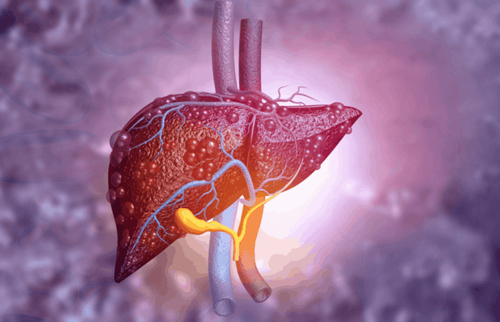
Tình trạng xơ gan do nhiều nguyên nhân khác nhau gây ra
2. What are the means to diagnose cirrhosis?
In addition to the physical examination of the above findings, the physician may palpate the liver indirectly through the abdominal wall, assessing its size, density, structure, and surface. A fibrous liver is often atrophied, rough, rough, and has a rough, irregular surface instead of a smooth, smooth surface.In addition, to accurately assess liver structure as well as liver function, some of the following cirrhosis tests should be ordered:
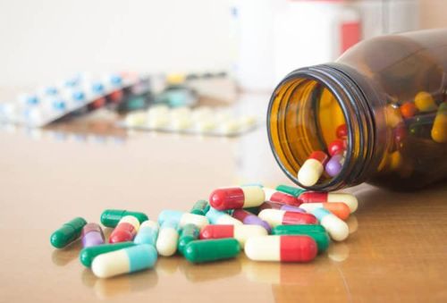
Blood tests: Cirrhosis will cause the platelet count to decrease, anemia, coagulation dysfunction, decreased protein synthesis in the blood, increased liver enzymes if cirrhosis is caused by liver damage... Imaging: The morphology of the liver will be observed first by the abdominal ultrasound tool. The liver structure is dense, full of fibrous tissue, which is a sign of cirrhosis. Some other imaging tools can be used when more in-depth investigation is needed, such as computed tomography to find liver tumors and stones in the liver. Measurement of liver elasticity: Because when the liver has cirrhosis, the liver parenchyma contains a lot of neoplastic fibrosis, the elasticity of the liver tissue will decrease. Thereby, the assessment of liver elasticity by ultrasound will help to detect liver fibrosis early as well as the stage of cirrhosis. Endoscopy: With a thin, flexible tube with a light and camera attached at the end of the tube inserted into the mouth, the surface of the esophagus and stomach shows signs of large varicose veins that point to cirrhosis. . Biopsy: This is an invasive but “gold standard” method of diagnosing cirrhosis. Therefore, this modality is only indicated when the diagnosis is difficult and with rare etiologies by obtaining a tissue sample through transdermal needle puncture of the liver. Surgery: In some cases, cirrhosis is diagnosed during surgery when the doctor can see the entire liver.

Xét nghiệm máu cũng có thể giúp ích trong việc chẩn đoán xơ gan
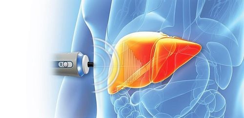
3. What is the diagnosis of cirrhosis by the Fibroscan method?
Fibroscan or hepatic elastography is a non-invasive test that helps assess the health of the liver in general and the presence and density of fibrous tissue in the liver parenchyma in particular. By using ultrasound technology to determine the extent of fibrosis or scarring that may be present in the liver, a diagnosis of cirrhosis can be confirmed or ruled out, denying that the above symptoms may be caused by disorders in other organ systems.FibroScan test results are always used in conjunction with other clinical data, laboratory results and liver imaging in individual patient management, personalization and optimization of treatment plans, and in long-term follow-up. For some patients, ultrasound elastography can even replace a liver biopsy.
Although elastography is a completely non-invasive, simple, and painless test that takes about 10 minutes to complete, this test has limitations in patients who already have effusion abdominal cavity, acute hepatitis, right heart failure, severe obesity, narrow intercostal space or inability to lie flat...
4. How is the procedure to diagnose cirrhosis by Fibroscan method?
4.1 Preparations to perform The patient is asked to wear loose clothing and to be able to lie flat on the table for 10 minutes or more without any problems.The patient was instructed not to eat or drink any food for at least 3 hours before the test to increase the reliability of the test.
The liver elastography scan usually takes 10 to 15 minutes to complete. However, patients should be present 30 minutes in advance to allow time to prepare.
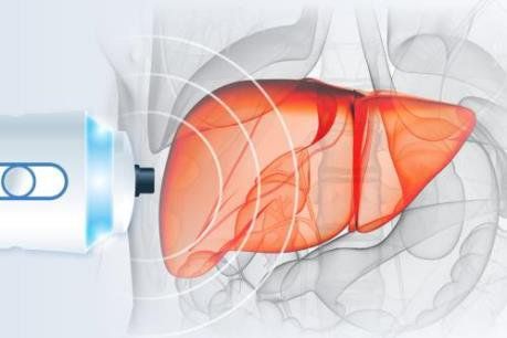
Quy trình thực hiện chẩn đoán xơ gan bằng phương pháp Fibroscan có thể kéo dài 15 phút
The technician will place the FibroScan probe between the ribs on the right side of the lower chest wall. A series of ultrasonic pulses are emitted and the echo is recorded on the device, calculating an overall liver stiffness score.
This score is then assessed by a hepatologist to predict the likelihood of cirrhosis or advanced cirrhosis.
4.3 End of procedure The patient can go home and resume normal activities.
Results will be sent back to the appointed doctor in about one to two weeks.
5. Advantages when diagnosing cirrhosis by Fibroscan method
Fibroscan or hepatic elastography is a simple, painless, noninvasive test used to make a relatively accurate assessment of the health of the liver. Therefore, liver elastography has gradually replaced most of the traditional indications for liver biopsy.By using a combination of elastic waves generated by mechanical pulse and ultrasound technology, liver elasticity helps to evaluate the stiffness of the liver. If present, this is evidence of histological level, detecting early signs of liver damage, presence of liver fibrosis, which can eventually lead to cirrhosis and liver cancer.
From there, the results of the elastography of the liver will decide the treatment plan and follow up the patient in the future. If severe liver damage is found, the patient will need to see a liver specialist to determine the cause of the disease and then have appropriate management. In the absence of cirrhosis or with only mild fibrosis, modification of lifestyle interventions is often sufficient to prevent the development of actual liver disease as well as its progression.

Kết quả siêu âm độ đàn hồi mô gan
In summary, the diagnosis of cirrhosis by Fibroscan method is a new, non-invasive, rapid and reparable method to evaluate liver fibrosis by measuring liver stiffness. From there, patients will reduce the risk of having to have a liver biopsy as well as detect the disease early and treat it properly.
Currently Vinmec International General Hospital provides services to diagnose cirrhosis by Fibroscan method. At Vinmec, the ultrasound-elastic technique of liver tissue is performed with the most modern equipment today, including:
LOGIQE 9 machine: a powerful and flexible ultrasound imaging system that meets a wide range of needs. General examination, with advanced, intuitive capabilities. LOGIQE 9 allows you to use professional tools to obtain excellent images and facilitate a smooth examination. The new Shear Wave technology is used to qualitatively and quantitatively measure the elastic energy of the parenchyma. Through color coding and the value of elastic levels in pressure units kPa. The Shear Wave parenchymal elastography tool allows the user to actively localize the survey right on the ultrasound image. Images obtained during the survey can be easily marked and tracked. In addition, the hospital also has the participation and companionship of leading domestic and foreign experts, rich in professional experience and best customer care services, helping customers feel completely secure when visiting. .
Before accepting a job at Vinmec Central Park International General Hospital, the position of Doctor of Radiology since February 2018, Doctor Trinh Le Hong Minh used to work as a resident in the Department of Radiology at other hospitals. Hospitals: Cho Ray, University of Medicine and Pharmacy, Oncology, People's Gia Dinh, Trung Vuong... from 2012-2015. Officially worked at Cho Ray Hospital from 2015-2016, City International Hospital from 2016-2018.
To register for examination and treatment at Vinmec International General Hospital, you can contact the nationwide Vinmec Health System Hotline, or register online HERE.
References: hepmag.com , mskcc.org , ncbi.nlm.nih.gov , nhs.uk