This is an automatically translated article.
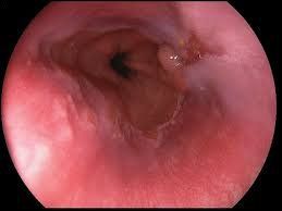
The article was written by MSc Mai Vien Phuong - Department of Examination & Internal Medicine - Vinmec Central Park International General HospitalEosinophilic esophagitis is an emerging disease and was once considered a rare but increasingly common condition, due to abnormal infiltration of eosinophils into the esophageal mucosa, affecting function. Esophageal structures—may progress to decrease in diameter and narrowing of the esophagus. Careful evaluation of patients with dysphagia is necessary to detect the disease and initiate early treatment.
1. White blood cell classification and function
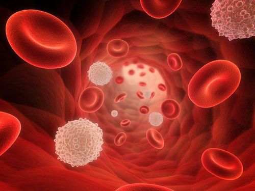
There are three main types of white blood cells: granulocytes, lymphocytes, and monocytes. Granulocytes are white blood cells with small protein-containing granules. There are three types of granulocytes:Basophils: They make up less than 1% of the white blood cells in the body and usually appear in increased numbers after an allergic reaction. Eosinophils: These are white blood cells responsible for responding to infections caused by parasites. Neutrophils: These represent the majority of white blood cells in the body. They act as “garbage collectors”, helping to surround and destroy bacteria and fungi that may be present in the body.
2. What are eosinophils?
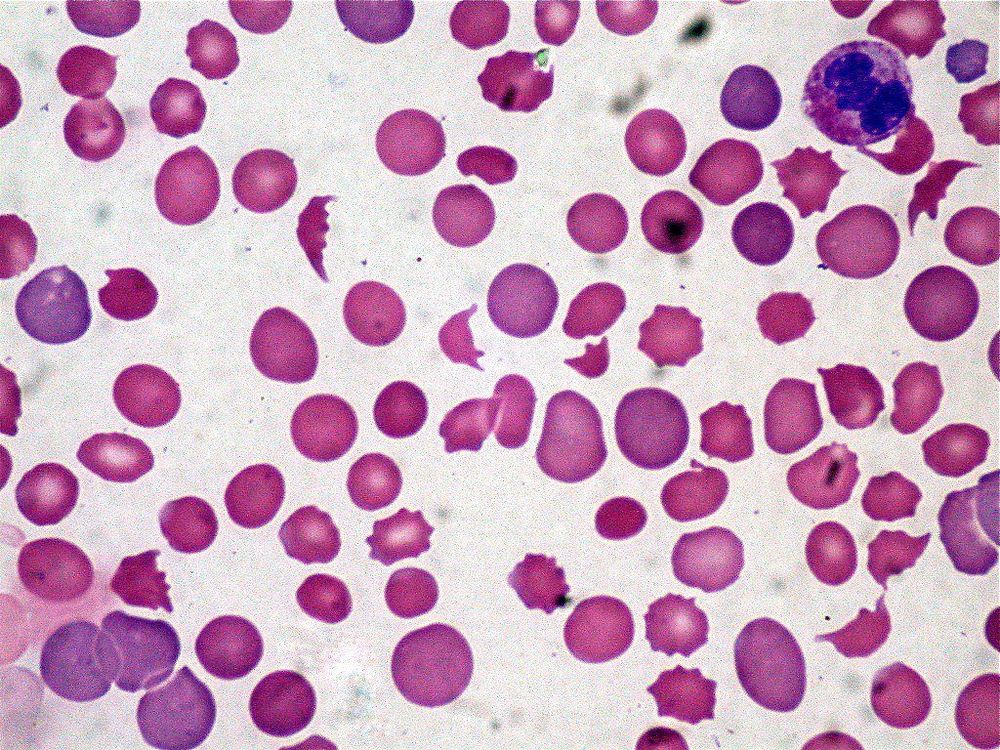
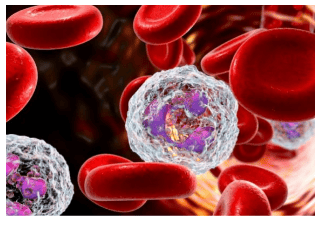
Therefore, large quantities of eosin can accumulate in tissues such as the esophagus, stomach, small intestine and sometimes in the blood when such individuals are exposed to the allergen. The allergens that cause this EE esophagitis are not known, only known that they enter through inhalation or ingestion.
Eosinophils in particular and types of white blood cells in general have the following characteristics:
Ability to change shape, penetrate the cell wall to move to places where white blood cells are needed. Capable of moving with a prosthetic leg (amoeba-like) at a speed of 40 mm/min. When there are substances produced by inflammatory tissue or produced by bacteria or there are chemicals introduced from the outside into the body, attracting eosinophils or driving them away. Phagocytosis: Capable of swallowing foreign objects that enter the body.

Although eosinophils are part of the immune system, their response is not always good for the body. Sometimes, they are part of conditions such as food allergies, inflammation in the body's tissues.
3. What is the esophagus? Where is the esophagus located?
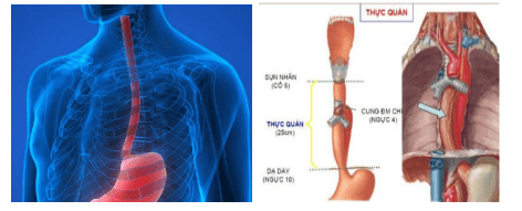
The esophagus is the first part of the digestive tube that carries food from the pharynx to the stomach, about 25cm long, relatively mobile, attached to surrounding organs by loose structures, flattened in shape because the walls are close to them. each other and have a tubular shape when swallowing food.
Above the esophagus, it connects to the pharynx at the level of the 6th cervical vertebrae, below the gastric outlet at the cardia, at the level of the 10th thoracic vertebrae. The esophagus is located behind the trachea, descends to the posterior mediastinum, is located posteriorly. posterior to the heart and anterior to the thoracic aorta, through the diaphragm into the abdomen, to the stomach.
4. What is esophagitis?
Esophagitis is a condition in which the lining of the esophagus is damaged and leads to inflammation. Esophagitis has several causes: the most common cause is acid reflux, which leads to a burning sensation in the throat, although acid reflux can cause ulcers on the inner lining of the esophagus.
Other, rarer causes are viruses (herpes simplex), fungi (candida), drugs (tetracycline), radiation therapy (lung cancer treatment). Physicians believe that eosinophilic esophagitis is a type of esophagitis caused by asthma-like allergies, or fever, allergic rhinitis and atopic dermatitis, and even some other allergens.
5. What is eosinophilic esophagitis?
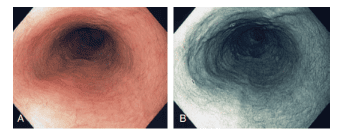
Eosinophilic esophagitis (VTQDBCAT) is an eosinophilic infiltration of the esophagus that causes clinical symptoms.
6. How is eosinophilic esophagitis found?
This is a new disease that has only really received attention in the last 20 years. The first case described by Landres in the literature in 1978 was a case where a patient was initially diagnosed with achalasia and then discovered a thickening of the muscular layer in the esophagus and histopathology showed There was an eosinophilic infiltrate.
During the period from 1989 to 1994, a few more studies reported a series of cases that reported cases with dysphagia but no obstruction, normal esophageal pH and at birth. The number of eosinophils in the esophageal mucosa was found to be increased.
In 1995, Kelly et al. reported a number of pediatric patients with eosinophilic esophageal infiltrates who did not respond to reflux treatments including medication and Nissen surgery but achieved relief with strict dietary regimens. Strictly include amino acids according to the formula.
7. Is eosinophilic esophagitis common?
The incidence of VTQD varies by region, in which the incidence rate as recommended by the European Gastroenterology Society in 2017 ranges from 13-49/100,000 population and the annual incidence ranges from 1-20/100,000. people. The prevalence of symptomatic adult oesophagoscopy during upper gastrointestinal endoscopy is 7% and this incidence may increase to 23% in patients with dysphagia and 50% in patients with dysphagia. choking on food. In Asia, Japan was the first country to report a case in 2006 and then more and more cases were recorded in some other countries...According to a review study of VTQDBCAT tickets in Asia, from 2005. 1980 to 2015, 25 studies with 217 patients were reported in Japan, Korea, China and Saudi Arabia.
Epidemiological, clinical and histopathological studies all show a higher incidence in men and a male:female ratio of 3:1. Studies that focus on typical symptoms of VTQDBCAT, which are difficulty swallowing and choking with food, also show a male predominance.
Median age of diagnosis in adults aged 30 to 50 years and age at detection was not associated with progression of symptoms. However, some studies show that the average time from the onset of symptoms to diagnosis is quite late, from 4.2 to 4.8 years.
8.Eosinophilia in the esophagus can be seen in what diseases?
Normally, the lining of the esophagus may have some lymphocytes but no eosinophils. In the presence of allergic inflammation, the epithelial cells of the mucosal layer hyperplasia and the appearance of eosinophils begins. The presence of eosinophils in the esophageal epithelium is called esophageal eosinophilia. However, based on histopathological results alone, the diagnosis of VTQDBCAT cannot be confirmed. This condition can also be seen in other diseases of the esophagus such as gastroesophageal reflux disease (GERD), eosinophilia in response to proton pump inhibitors (PPI REE). ), cardiac spasm or some extra-esophageal diseases such as Celiac disease, Crohn's disease, infection, eosinophilic syndrome, drug sensitivity, vasculitis, pemphigus, connective tissue diseases, waste transplant.... In 2007, the first consensus in the world on VTQDBCAT did not clearly state the definition of this pathology, but in the second consensus released in 2011, VTQDBCAT was defined as a chronic esophageal disease. immunological/antigenic disease characterized by symptoms associated with esophageal dysfunction and inflammation with the predominant presence of eosinophils on histopathology. This definition is still agreed upon in the follow-up recommendations of the American Gastroenterology Society 2013 and the European Gastroenterology Society 2017.
Please dial HOTLINE for more information or register for an appointment HERE. Download MyVinmec app to make appointments faster and to manage your bookings easily.


