This is an automatically translated article.
Cerebrospinal fluid leak in the skull base is a rare condition and difficult to intervene surgically. However, until recent times, with the advancement of endoscopic techniques, this condition can be performed in a less invasive way but still ensure effective treatment, and the postoperative process is also effective. has become simpler. Since then, the patient's life has also been significantly improved, significantly reducing the risks related to cerebrospinal fluid leakage that can cause.
1. Introduction to endoscopic surgery to treat cerebrospinal fluid leaks at the base of the skull
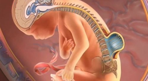
CSF leak is an abnormal flow of cerebrospinal fluid outside the meningeal cavity. The causes of the disease may be trauma, accident or surgery, tumor of the nasal cavity, or it may be congenital. Cerebrospinal fluid leaks of any cause are very rare and difficult to treat. In addition, the cases of cerebrospinal fluid leakage at the base of the skull can also be related differently to brain herniation and accompanying meningeal hernia, further complicating the management, especially when there is a meningeal herniation. big brain.
Previously, the treatment of cerebrospinal fluid leaks at the base of the skull often required open surgery with a frontal sinus flap. However, since the advent of endoscopic techniques and tools and continued development, endoscopic surgery for CSF leakage makes the intervention less invasive, allowing the Post-surgery simpler.
2. Indication and selection of patients to perform endoscopic surgery for CSF leakage in the skull base
The preoperative process for endoscopic CSF leak surgery includes a thorough clinical examination, nasal endoscopy, and radiological evaluation of the skull.
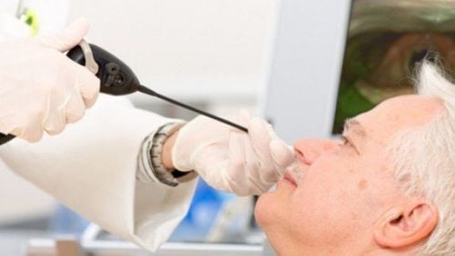
Endoscopic examination: Endoscopic examination can sometimes reveal a large meningioma. At the same time, nasal endoscopy can also identify any local anatomical variations that may interfere with surgical manipulation such as septal deviation or septal spines, synapses,...
Evaluation of images Image: High resolution computed tomography and magnetic resonance encephalopathy are essential to diagnose and locate the CSF leak as well as guide the intervention protocol.

Người bệnh cần được nội soi mũi trước khi phẫu thuật nội soi điều trị rò dịch não tủy nền sọ
3. How is endoscopic surgery for cerebrospinal fluid leak performed?
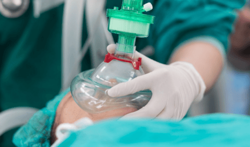
Endoscopic CSF leak surgery is performed under general anesthesia and the patient is placed in the supine position.
The entire intervention can be performed by one surgeon or by two surgeons using the 3 or 4 hand technique. The specific selection of the most appropriate surgical approach will depend on the location and size of the cerebrospinal fluid leak as well as the available equipment and the skill and experience of the surgeon. In which, thorough knowledge of frontal sinus anatomy, endoscopic skills in accessing the skull base area are essential to be able to fully approach the acquired defect.
Endoscopes, consisting of conduction lenses into a monitor allowing the entire surgical field to be viewed from the outside and interventional devices, are inserted into the nostrils and access the base of the skull. By carefully avoiding trauma to the olfactory bulb as well as to all anterior neurons around the frontal sinus, the floor bone fragment is exposed and must be opened to access the cerebrospinal fluid leak. If the instruments cannot reach the defect site after complete opening of the sinus cavity, a ethmoidectomy should be performed. The extent of the sinus opening must be adapted to the type of cerebrospinal fluid leak and it is not always necessary to have a large sinusectomy to intervene. It is the advantage of laparoscopy that overcomes the limitation of surgical field vision in these circumstances.
When the defect or meninges are exposed and the surgeon sees on the screen, a 5mm strip of healthy mucosa is removed around the defect to ensure better graft adhesion and promote healing. osteogenesis around the defect. Simultaneously, a piece of meninges is pulled out and tightened into the dura. If needed, the surgeon can remove the excess meninges. From there, the defect site causing the cerebrospinal fluid leak in the skull base will be sealed. In the case of spontaneous CSF leakage, the bone wall surrounding the lacunar is usually very thin and must be handled with great care to avoid unintentional enlargement of the lacunar.
Finally, the surgeon will meticulously remove the excess mucosa around the defect, closing the interventional skull base layer by layer but still ensuring ventilation for the sinuses. The endoscopic instruments will be removed and the patient will be monitored continuously until recovery.
4. Care after endoscopic surgery for cerebrospinal fluid leak
Although no consensus has been reached on the need for antibiotics after endoscopic CSF leak surgery, most surgeons nevertheless use intravenous antibiotics that are active against cocci. Gram-positive for 24 to 48 hours after the intervention to prevent infection due to surgery in general as well as infection of the central nervous system - a complication of cerebrospinal fluid leak in particular. Besides, pain relievers and laxatives are also prescribed.
The recommended period of strict bed rest after endoscopic CSF leak surgery varies considerably from case to case as well as the course of the intervention, typically ranging from 1 day to 2 weeks, and is largely dependent on the patient's condition. depends on the risk of recurrent cerebrospinal fluid leaks. After going home, the patient should be instructed and advised to regularly perform maneuvers to unclog the nasal cavities to avoid prolonged crusting and fluid accumulation. At the same time, the patient must avoid lifting heavy objects and blowing their nose or doing other activities that increase the pressure on the base of the skull.

Sau phẫu thuật nội soi rò dịch não tủy, người bệnh được dùng thuốc theo y lệnh
5. Complications of endoscopic cerebrospinal fluid leak surgery
Complications of endoscopic CSF leak repair that have been documented in studies are meningitis, brain abscess, subdural hematoma, olfactory dysfunction, and headache.
In which, the typical acute complications for endoscopic surgery with access to the skull base are trauma to the anterior nasal artery that can cause immediate or late postoperative nosebleeds, or even hematomas that need to be resolved. emergency pressure. A specific feature of the cranial base access route through the nostrils is that it is sometimes very narrow, leading to a high risk of medium- or long-term postoperative stenosis and, consequently, of headache and chronic sinusitis.
In summary, endoscopic surgery for cerebrospinal fluid leakage, thanks to advances in endoscopic techniques, has achieved many outstanding results compared with previous open surgery for the treatment of CSF leaks in general. However, compared with other endoscopic interventions, because the surgical field of cerebrospinal fluid fistula is sometimes limited, the success of the operation depends not only on the preoperative imaging assessment but also on the knowledge of anatomy, skills of surgeons as well as post-operative care.
If you have a need for consultation and examination at Vinmec Hospitals under the national health system, please book an appointment on the website (vinmec.com) for service.
Please dial HOTLINE for more information or register for an appointment HERE. Download MyVinmec app to make appointments faster and to manage your bookings easily.


