This is an automatically translated article.
Digitalization of the background and occlusion of gastric varices was evaluated successfully when the entire varicose veins of the stomach collapsed due to fibrosis, the patient was no longer enhanced through CT scan images and other The branch has no flow.
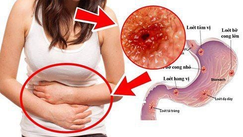
1. What is gastric varices?
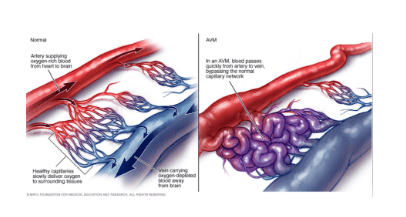
Gastric varices is a condition in which the veins in the stomach are dilated. When normal blood flow to the liver is blocked by a blood clot or there is scarring in the liver, blood flows into smaller blood vessels. However, small blood vessels are not responsible for transporting a large volume of blood, leading to overload.
Rupture due to hemorrhagic varices usually occurs in the esophageal veins and vena cava system. When that happens, blood vessels can leak or even burst. The red spots in the large dilated veins are the place at greatest risk of rupture, if too much blood is lost, the patient will go into shock and die.
2. Causes of gastric varicose veins
Gastric varices are one of the common complications in people with portal hypertension syndrome. The following causes are also one of the factors that lead to this disease:
Cirrhosis: Cirrhosis causes the flow of blood in the portal vein (which is responsible for transporting blood from the stomach and intestines to the liver) prevent. Therefore, blood will find the small veins to move. However, small blood vessels with thin walls, when subjected to high pressure, will bulge, and eventually burst, causing bleeding. Blocked blood clot: Blood will not be transported if a clot forms in the portal vein. Parasitic infections: Schistosomiasis is a parasite that damages the liver, lungs, intestines, and bladder. This leads to gastric varices.

Giãn tĩnh mạch dạ dày là tình trạng các tĩnh mạch ở dạ dày bị giãn ra.
3. Symptoms of gastric varicose veins
Gastrointestinal varices have no obvious clinical manifestations until the patient has bleeding. When bleeding, patients will appear the following common symptoms:
Vomiting blood; Pass black stools ; Dizziness of mind; Loss of consciousness; For people with liver disease, there will be additional symptoms such as: Jaundice, easy bruising, bleeding, yellow eyes, ascites ...
4. Digital scan to erase background and plug of gastric varicose veins
4.1. What is a digital scan to erase the background and occlusion of gastric varices?
Digital background erasing technique is an X-ray angiogram system, which can intervene intravenously when using a catheter with a balloon, to position the left gastro-renal vein. .
Before occlusion of varicose veins, the technician will inflate the balloon to block the flow and avoid reflux. Then, embolize the communication channels of the varicose veins with the inferior vena cava system.
4.2. Advantages of digital imaging to erase the background and occlusion of gastric varices
Digital angiography to clear the background is widely applied in the diagnosis of gastric varices occlusion. The advantages of this technique are:
Minimal intrusion; Safety for patients; Prevention of high risk of recurrence in gastric varices.
4.3. Contraindications to digital scan to clear the background and occlusion of gastric varices
If the patient has the following signs and pathologies, it is not possible to use digital imaging techniques to erase the background and occlude gastric varices:
People who are allergic to contrast agents; Have renal failure with serum creatinine level > 1.5 mg/dl; Portal vein occlusion, ascites; Existing esophageal varices; Pregnant women.

Chụp số mạch hóa xóa nền được áp dụng rộng rãi trong chẩn đoán nút tắc búi giãn tĩnh mạch dạ dày.
4.4. Digital imaging process to erase the background and occlusion of gastric varices
To conduct digital scan to erase the background and plug gastric varices, patients need to fast for 6 hours before the procedure, can drink water but not more than 50 ml. The procedure is as follows:
Step 1: Perform local anesthesia, make skin incisions, poke into the lumen to take angiograms to assess the damage; Step 2: Insert the balloon catheter into the gastro-renal vein. Inflate the balloon to block the flow of this venous shunt; Step 3: Insert the catheter into the varicose vein through the balloon catheter. After that, sclerotherapy and embolization are injected into the varicose veins through the microcatheter and the microcatheter is locked, the catheter has a balloon to prevent the fibrous material from regurgitating; Step 4: Take the patient to the recovery room, the catheter and microcatheter are locked but still in the artery. After 4-24 hours, abdominal computed tomography with contrast injection to evaluate the degree of obstruction of gastric varices; Step 5: Check the degree of occlusion by taking a pulse again. Step 6: Withdraw catheter and microcatheter. Step 7: Close the vascular access to end the procedure. Digital erasure and occlusion of gastric varices were evaluated successfully when the entire dilated gastric varices collapsed due to fibrosis, the patient no longer enhanced through CT scan images and branches. there is no more flow.
In summary, varicose vein occlusion carries a high risk of rupture, resulting in massive blood loss, shock, and death. Therefore, patients need to go to a reputable hospital to conduct examination and treatment as soon as there are signs of varicose veins. Currently, Vinmec International General Hospital is one of the top quality hospitals in the country, trusted by a large number of patients for medical examination and treatment. Not only the physical system, modern equipment: 6 ultrasound rooms; 4 DR X-ray rooms (1 full-axis, 1 intensifier, 1 synthesizer and 1 mammogram); 2 portable X-ray machines DR; 2 multi-row computer tomography rooms with receivers (1 128-series machine and 1 16-row machine); 2 rooms for magnetic resonance imaging (1 machine 3 Tesla and 1 machine 1.5 Tesla); 1 room for 2 levels of interventional angiography and 1 room to measure bone mineral density.... Vinmec is also the place where a team of experienced doctors and nurses will gather a lot of support in diagnosis and detection. early signs of abnormality in the patient's body. In particular, with a space designed according to 5-star hotel standards, Vinmec ensures to bring the patient the most comfort, friendliness and peace of mind.
Please dial HOTLINE for more information or register for an appointment HERE. Download MyVinmec app to make appointments faster and to manage your bookings easily.


