This is an automatically translated article.
The article was professionally consulted by Specialist Doctor I Tran Cong Trinh - Radiologist - Radiology Department - Vinmec Central Park International General Hospital.
Pump occlusion of peripheral vascular malformations under the guidance of digital background erasure is a method of treatment for peripheral vascular malformations to better evaluate the lesions and vascular pathology before indication for intervention. circuit.
1. Concept of angiography and direct occlusion of peripheral vascular malformation
Percutaneous angiography and direct percutaneous peripheral embolization are techniques indicated in the treatment of peripheral vascular malformations, which can be applied alone or in combination with traditional embolization techniques. This method is performed by inserting a needle into the malformation, then conducting contrast angiography to review the hemodynamic status of the lesion. Finally, embolize with drugs.
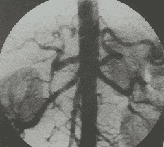
The purpose of digital erasure scan (DSA scan) in peripheral vascular malformation technique is to study the blood vessels in the body to be able to better assess the lesions and vascular pathology before making a decision. vascular intervention.
The purpose of digital erasure scan (DSA scan) in peripheral vascular malformation technique is to study the blood vessels in the body to be able to better assess the lesions and vascular pathology before making a decision. vascular intervention.
2. Indications and contraindications to pump occlusion of peripheral vascular malformation
2.1. Point
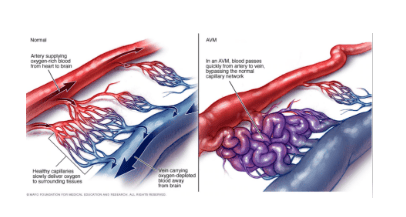
Patients with arteriovenous malformations Patients with venous malformations Hemangiomas.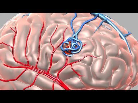
Bệnh nhân dị dạng động tĩnh mạch được chỉ định thực hiện
2.2. Contraindications
The area intended for direct puncture is showing symptoms of inflammation, infection, skin and soft tissue necrosis; Reactions to iodinated contrast agents; Signs of clotting disorder: prothrombin less than 60%, INR > 1.5, platelet count < 50 G/l.3. Procedures for angiography and occlusion of peripheral vascular malformations
3.1. Open the way to the heart
Perform antiseptic, local anesthetic at the target skin area, conduct skin incision technique; Use a needle of appropriate size to poke the lesion; This step can be performed under ultrasound guidance and/or a DSA scan.3.2. Angiography to assess injury status
Connect the puncture needle to the extension cord; Conduct angiography to study and evaluate the hemodynamic status of the lesion and adjacent vessels.3.3. Treatment intervention
Select materials that cause embolism (metal helix, bio-glue (nBCA, Onyx), fibrous agent (Tromboject) or Ethanol). The selection depends on the morphological characteristics and hemodynamic properties of the lesion; Insert embolization material into the injury site to embolize the vessel; In case the embolization material is a liquid solution (bio-glue) and the lesion has a large flow rate and has drainage veins, it is necessary to combine the placement of an intravenous Garo above the lesion (proximal limb). .3.4. Post-intervention assessment
Angiography to evaluate circulation after revascularization of the lesion. Close the vascular access and complete the procedure.4. Evaluation of results
The results of the peripheral vascular malformation pump procedure are considered successful when all the malformations are removed from the circulation and there is no flow signal.In addition, the arterial branches supplying blood to the downstream area and draining veins are still able to circulate normally.
Bơm tắc dị dạng mạch ngoại biên có thể gây viêm da hoại tử do thiếu máu tại chỗ
5. Risk of accidents and how to handle them
Depending on the selected embolization material and the condition of the lesion, the patient may sometimes experience a number of different complications:Hematoma at the opening site: Carrying out a pressure dressing to stop bleeding and follow; Distal embolism: This case is usually because the embolized material is a metal spiral, the flow is large, leading to the drift of embolized material down the extremities. Management strategies are offered depending on the extent of the embolism, but usually medical therapy is the only option. Necrotic dermatitis due to local ischemia: Commonly occurs with embolized material Ethanol, bio-glue due to local embolism. Management is usually medical treatment and local care. However, if necrotizing dermatitis spreads, abscesses, it is necessary to consult a specialist (dermatology, surgery); Contrast drug allergy: Perform the diagnosis and management of adverse events for contrast agent allergy. The method of pumping occlusion of peripheral vascular malformations is applied together with digital imaging to erase the background to better assess the lesions and vascular pathology before vascular intervention is indicated.
Vinmec International General Hospital has applied digital imaging technology to erase the background in examination and diagnosis of many diseases. Accordingly, the dsa imaging technique at Vinmec is carried out methodically and in accordance with the process standards by a team of highly skilled medical professionals and modern machinery. Thereby giving accurate results, contributing significantly to the identification of the disease and the stage of the disease and timely treatment.
Before taking a job at Vinmec Central Park International General Hospital, the position of Doctor of Radiology from September 2017, Doctor Tran Cong Trinh worked at Gia Dinh People's Hospital since 2007. -2017. In his role, Dr. Tran Cong Trinh has participated in guiding the teaching of students, residents, specialists and new doctors to the department


