This is an automatically translated article.
The article was professionally consulted by Specialist Doctor I Tran Cong Trinh - Radiologist - Radiology Department - Vinmec Central Park International General Hospital.
The treatment of venous malformation - a type of vascular malformation is usually done in a variety of ways. Direct sclerotherapy is a method that is being widely applied today.
1. Venous malformation and clinical signs of venous malformation
Venous malformations are among the most common, accounting for up to 50% of all vascular malformations. Accordingly, the clinical signs of venous malformation usually appear as a blue mass of the skin or mucous membranes. Venous malformation is more or less raised or as a superficial network of veins. When the lesion is soft, it can be easily deflated and inflated again on release, increasing volume in the downhill position and on exertion. Occasionally, calcified nodules may be palpable (known as venous stones).
Venous malformations are always present at birth, but may go unnoticed and begin to show symptoms during childhood or adolescence. Lesions may be small, localized, and unaffected for a long time. However, when it grows into muscles and organs and invades many anatomical structures, it can lead to severe functional or cosmetic distortions.
2. Treatment of venous malformations by direct sclerotherapy under digital angiography erasing the background
2.1. What are the treatments like?
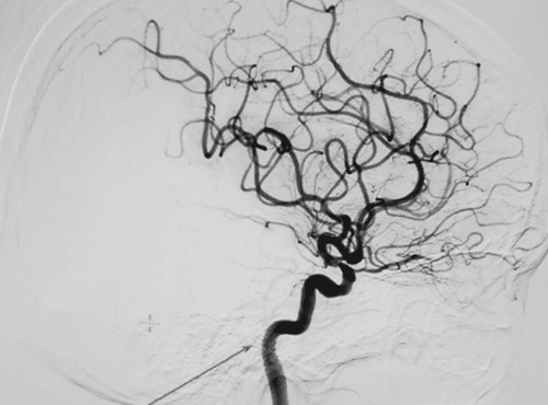
This method takes angiography and causes embolization directly through the skin. That is, it is performed by inserting a needle into the venous malformation, then angiography with contrast agent to evaluate the hemodynamic status of the lesion and finally, injecting the embolization drug.
This is an interventional method to treat peripheral vascular malformations, which can be applied alone or in combination with traditional embolization techniques.
The method is contraindicated in patients with the following problems:
Inflammation, infection, necrosis of the skin and soft tissues of the area intended for direct puncture Iodine contrast allergy Severe coagulopathy and loss of control (Prothrombin <60%, INR > 1.5, platelet count < 50 G/l) In the rest, patients with low flow can completely use this method to treat static malformations circuit.
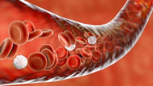
Bệnh nhân có rối loạn đông máu chống chỉ định thực hiện
2.2. How to proceed to treat venous malformations by sclerotherapy directly under digital angiography erasing the background
Open the way into the artery The doctor will give local anesthesia to the patient, then use a needle of suitable size to poke the lesion. This process can take place under ultrasound guidance and/or DSA imaging. Next - take an angiogram to assess the lesion and then connect the puncture needle to the extension cord.
Conduct a systemic angiogram to assess the hemodynamic status of the lesion and surrounding vessels.
Treatment intervention The doctor will monitor and depending on the morphological and hemodynamic characteristics of the lesion to decide on the choice of embolization materials: metal spiral (Coil), bio glue (nBCA). , Onyx), fiber (Thromboject) or Ethanol. After examining, the doctor inserts embolization material into the lesion to block the vessel.
In case the embolization material is a liquid solution (bio-glue) but the lesion has a large flow rate and has drainage veins, it is necessary to combine the Garo vein above the lesion (proximal limb).
Post-intervention evaluation The doctor will take angiography to assess the circulation after re-vibration and then close the way into the lumen, ending the procedure.
2.3. When is this treatment considered successful?
The results of this method are considered successful when all the deformities are removed from the circulation, and there is no flow signal. At the same time, the branches of the arteries supplying blood to the downstream area and the draining veins still circulate normally.
Phương pháp điều trị này có thể gây viêm da hoại tử do thiếu máu
3. Notes to know when using methods to treat venous malformations
Depending on the embolization material selected, this method can have different complications.
Distal embolism: This complication is common because the embolized material is a metal spiral, the flow is large, and the embolized material is pushed down the extremities. Depending on the degree of embolism, there is a treatment strategy. Usually, only medical treatment is needed. Necrotic dermatitis due to local ischemia: common for embolized materials is Ethanol, bio-glue due to local embolism. When this is the case, the patient needs medical treatment and local care. Need specialist consultation (dermatology, surgery) in case of diffuse necrotizing dermatitis, abscess.
Hematoma at the site of opening the way into the vessel lumen: The doctor will apply pressure to stop the bleeding. If you have an allergy to contrast agents: you can follow the anti-allergy/shock regimen to manage. Vascular malformation in general and venous malformation in particular is a disease that needs to be treated thoroughly. Direct sclerotherapy holds the promise of becoming a pioneer due to its amazingly high degree of success and the potential for faster and healthier recovery for patients.
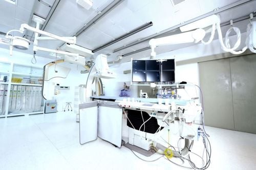
DSA background removal digital capture technique has been widely used. At Vinmec International General Hospital, there is a team of experienced specialists, along with modern equipment, using digital angiography to erase the background of DSA in the diagnosis and treatment of diseases, bringing the best results. optimal treatment results for customers.
For examination and treatment at Vinmec International General Hospital, please come directly to Vinmec Health System or register online HERE.


