This is an automatically translated article.
This article is professionally consulted by Master, Doctor Vu Huy Hoang - Radiologist - Department of Diagnostic Imaging and Nuclear Medicine - Vinmec Times City International Hospital. Doctor Vu Huy Hoang has 10 years of experience working in the field of diagnostic imaging.Digital erasure of the background and node of the portal vein system is a method of treatment for primary liver cancer when the patient has an indication for major hepatectomy but the remaining liver volume is not enough and there is no liver failure.
1. What is a digital background eraser and portal vein node?
Digital erasure of the background and portal vein node is a method of treating primary liver cancer when patients with liver cancer are indicated for major hepatectomy but the remaining liver volume is not enough and there is no liver failure.This method completely treats the tumor, but about 80% of cases when it is found that it is no longer possible to operate due to many reasons, one of the reasons that cannot be operated is the remaining liver volume after surgery. insufficient. The planned portal vein node surgery causes enlargement of the remaining liver, increasing the possibility of surgical treatment for the patient.
2. Indications and contraindications for digital background erasure and portal venous nodes
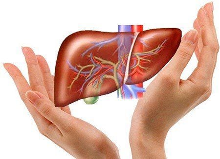
Patients with liver cancer are indicated for major liver resection but the remaining liver volume is not enough. No liver failure. Digital imaging is contraindicated to remove the background and nodes of the portal venous system when:
Liver tumor has extensive spread in the liver (lesions in subdivisions I, II, III) Left liver tumor (injury to subsegments VI and VII) Liver tumor with vascular invasion Liver tumor with biliary dilatation, portal hypertension. Patients with heart failure, severe kidney failure Have lymph node metastasis, lung metastasis Coagulation disorders, platelets Pregnant women.
3. Steps to take digital scan to remove background and node of portal venous system
4. Complications and how to handle after implementing the method
Intra-abdominal bleeding: Check the bleeding site and perform embolization to stop the bleeding. Wrong displacement of the stopper material: Reposition. After the procedure, there may be subcapsular hematoma, abdominal pain (treated with common analgesics). The danger of liver cancer has shown the importance of screening and early detection of the disease. Therefore, Vinmec International General Hospital has provided customers with a package of screening and early detection of liver cancer to screen for liver cancer pathology for people at high risk of disease such as: alcoholics, cirrhosis, family history of liver cancer, cirrhosis, hepatitis B virus infection, chronic hepatitis C,...Service selection Package for screening and early detection of liver cancer, patients will be examined, consulted and performed tests, diagnostic imaging to evaluate liver function, liver diseases and liver cancer screening. With a system of modern machinery and a team of highly qualified and experienced doctors, Vinmec is committed to the best protection for the health of patients.
Please dial HOTLINE for more information or register for an appointment HERE. Download MyVinmec app to make appointments faster and to manage your bookings easily.
SEE MORE
Prognosis of liver cancer treatment by stage The danger of liver cancer Why is liver cancer more common in men than in women?


