This is an automatically translated article.
1. Expression of hemorrhagic stroke
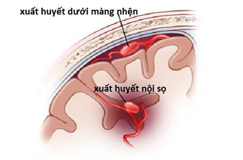
The typical clinical sign of a patient with a brain bleed is that the disease has a very sudden onset, with typical signs such as: headache, vomiting, or may be impaired consciousness (confusion, tumor, etc.) coma, coma), may be accompanied by motor paralysis, breathing disturbances, increased sputum secretion, especially disturbances of consciousness, which are common mainly in patients with large, deep hematomas near the midline. .Cases of blood spilling into the ventricles, based on the volume of blood flowing, a deep coma can occur with manifestations such as: convulsions, vomiting, brain loss, severe respiratory disorders, blood The pressure rises rapidly and then gradually decreases... Especially, most patients will die very soon, within the first 24 hours.
For milder cases, the coma may subside, after the patient comes out of the coma, most of the consciousness will be restored over time, but there may also be disturbances of consciousness featured.
Patients with small autologous hematomas will have a simpler and more favorable clinical course, neurological symptoms will be gradually restored and the hematoma will be self-absorbed, leaving very few dangerous sequelae.

2. Diagnosis of hemorrhagic stroke
2.1 CT brain scan CT brain scan is the main standard in diagnosing bleeding in the brain, CT scan of the brain also helps to assess how the brain bleeds progress in the first 6 hours.
According to research, within the first 7 days, around the hematoma, there will be an edema halo appearing, which is caused by hemoglobin degeneration and brain edema, along with damage to nerve cells, macrophages, neutrophil .
Then, from 7-14 days, the accumulated blood will gradually dissipate, from 15-22 days, the hematoma can completely co-densify with the patient's brain organization. And after 3 to 4 weeks, the hematoma will disappear, and leave the foci of liquefaction, on the CT scan, the focus will begin to decrease in density and be smaller than the old hematoma or have a sickle shape. , comma shape.
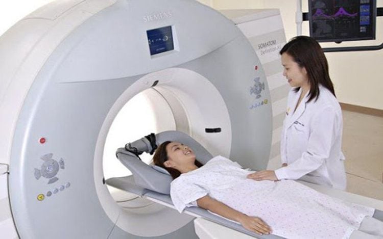
2.2 Cranial magnetic resonance imaging (MRI) Cranial magnetic resonance imaging (MRI) can also identify pathological images of the brain more clearly, especially longitudinal images of the brain, foci of lesions in the brain. brain stem or posterior fossa, vascular malformations, or occlusive stenosis are detected.
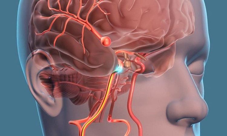
2.3 Digital subtraction angiography (DSA) Digital erasure angiography (DSA) is mostly indicated in the diagnosis and intervention of cerebral aneurysms, dural arteriovenous shunts, arteriovenous shunts. carotid artery in the cavernous sinus, arteriovenous malformations of cerebral vessels, stenosis and occlusion of the internal or extracranial carotid arteries, cerebral venous thrombosis, diseases of intracranial growths...
MRI system 3.0 Tesla at Vinmec hospitals across the country are equipped with state-of-the-art equipment by GE Healthcare (USA) with high image quality, allowing for a comprehensive assessment, not missing the injury, but also reducing the time taken to capture. Silent technology helps to reduce noise, create comfort and reduce stress for customers during shooting, resulting in better image quality and shorter shooting time.
With the state-of-the-art MRI system and the application of modern cerebral vascular intervention methods, a team of experienced and well-trained neuroradiologists and specialists, Vinmec is the address for risk examination and screening. Prestigious stroke engine trusted by customers.
Please dial HOTLINE for more information or register for an appointment HERE. Download MyVinmec app to make appointments faster and to manage your bookings easily.
3. Treatment of brain bleeding
According to the guidelines of the Vietnam Cardiology Association and the American Stroke Association, patients with cerebral hemorrhage will be treated at the Stroke Unit, the Emergency Department, the Stroke Center or the Intensive Care Unit. . Medical treatment: Parenchymal intracerebral hemorrhage due to hypertension is a medical emergency, so it should be treated quickly, promptly and comprehensively in all aspects according to the following principles:Complete treatment represent the patient in terms of neurological status and vital functions (ensure maintaining BP < 140/90 mmHg for patients in the first hours of admission, perform airway circulation...). Regular monitoring is required to prevent rapid progression of hematomas and to prevent and treat neurological complications if present (eg, interstitial edema, seizures), infections, ulcers. ..., at the same time to prevent relapse for patients, and restore function early. Management of BP: Brain bleeding fluctuates in the range of 150-220 mmHg, there are no contraindications to treatment, so lowering SBP ≤140 mmHg is safe for patients (Class I, level A) and helps to improve consequences functions. Monitoring and initial management, care under the guidance of medical staff with patients with brain bleeding. Treatment of blood glucose: Conduct monitoring along with adjustment of blood glucose in the patient's blood if increased or decreased. Treatment of fever: Patients with brain bleeding with signs of high fever must be treated as directed by medical staff. Timely diagnosis and treatment of hemorrhagic stroke plays an extremely important role in providing first aid and saving lives.
Currently, Magnetic Resonance Imaging - MRI/MRA is considered a "golden" tool for brain stroke screening. MRI is used to check the condition of most organs in the body, especially valuable in detailed imaging of the brain or spinal nerves. Due to the good contrast and resolution, MRI images allow to detect abnormalities hidden behind bone layers that are difficult to recognize with other imaging methods. MRI can give more accurate results than X-ray techniques (except for DSA angiography) in diagnosing brain diseases, cardiovascular diseases, strokes,... Moreover, the process MRI scans do not cause side effects like X-rays or computed tomography (CT) scans.\
Vinmec International General Hospital currently owns a 3.0 Tesla MRI system equipped with state-of-the-art equipment by GE. Healthcare (USA) with high image quality, allows comprehensive assessment, does not miss the injury but reduces the time taken to take pictures. Silent technology helps to reduce noise, create comfort and reduce stress for the client during the shooting process, resulting in better image quality and shorter imaging time. With the state-of-the-art MRI system With the application of modern methods of cerebral vascular intervention, a team of experienced and well-trained neurologists and radiologists, Vinmec is a prestigious address for stroke risk screening and screening. reliable goods.
In the past time; Vinmec has successfully treated many cases of stroke in a timely manner, leaving no sequelae: saving the life of a patient suffering from 2 consecutive strokes; Responding to foreign female tourists to escape the "death door" of a stroke;...
Please dial HOTLINE for more information or register for an appointment HERE. Download MyVinmec app to make appointments faster and to manage your bookings easily.


