This is an automatically translated article.
The article is professionally consulted by Master, Doctor Nguyen Viet Thu - Doctor of Radiology and Nuclear Medicine - Department of Diagnostic Imaging and Nuclear Medicine - Vinmec Times City International General Hospital.Computed tomography of the chest is an advanced imaging method that uses X-rays with greater intensity than X-ray films to examine the internal structures of the chest. This is a means with many advantages and gradually becomes an aid in the diagnosis, detection and monitoring of diseases in the thorax, heart, lungs and mediastinum. In addition, the doctor may prescribe contrast injection for chest CT scan when it is necessary to evaluate the absorption status of abnormal structures, thereby making more accurate judgments.
1. Overview of thoracic computed tomography
Computed tomography of the chest, also known as a chest CT scan, is an imaging technique that uses high-intensity X-rays directly into the chest. The resulting images are the result of electronic processing carried out through a pre-installed computer system with good image quality and detailed information. Chest computed tomography was first discovered in 1972 by physician Allan Cormack and physicist Godfrey Hounsfield. Since then, thoracic CT scan has been increasingly widely indicated and gradually replaced chest X-ray film in a number of diseases.Computed tomography of the chest has a relatively fast implementation time and provides detailed image quality when evaluating lesions in the chest wall, ribs, pleura, lungs, pulmonary vascular system, heart and other organs. organs inside the mediastinum. Computed tomography of the chest has outstanding advantages in detecting diseases or lesions that are missed on chest x-rays due to overlapping organs. Primary malignancies in the chest or metastases from other organs in the body, acute cases such as arterial dissection, bulging aneurysms can also be well investigated by this method. computed tomography of the chest.
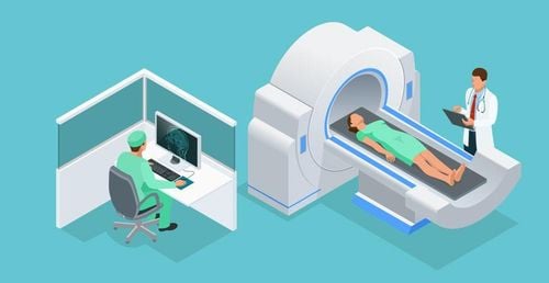
Chụp cắt lớp vi tính lồng ngực giúp phát hiện các tổn thương tại thành ngực
2. Indications and contraindications of thoracic computed tomography
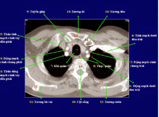
The doctor appoints the method of computed tomography of the chest when he wants to examine and evaluate abnormalities related to the internal structures of the chest, including the ribs, chest wall, lung parenchyma, pleura, vascular system. pulmonary blood, heart and great arteries, and structures within the mediastinum. Some situations that may require a CT scan of the chest are commonly seen in clinical practice such as:● The patient has a dull, persistent, unexplained chest pain or pain in the entire rib cage.
● When suffering from malignancies in other organs in the body such as the digestive system, liver, patients are often assigned a chest computed tomography scan to detect metastases, if any.
● After closed chest trauma, large bruises appear in the chest due to traffic accidents, work accidents and daily life.
Open chest trauma caused by bullets or knives
Assess the state of compression when there are tumors in the chest

Chụp cắt lớp vi tính lồng ngực được chỉ định cho người bị đau ngực không rõ nguyên nhân
People with a history of contrast allergy should not be indicated for contrast injection. chest.
Women who are suspected of being pregnant or are pregnant in the first trimester because X-rays have a risk of causing birth defects in the fetus
People with chronic kidney disease, kidney failure should not use contrast agents during a chest CT scan.
3. What preparation is required before conducting a chest computed tomography scan?
To ensure safety and effectiveness, patients should remember and adhere to a few notes before performing a thoracic computed tomography scan:Need to fast for at least 4 hours before starting the procedure. perform technique
● Immediately notify a medical staff if you suspect that you are pregnant or are pregnant. Many studies have shown that X-ray exposure during the first trimester of pregnancy increases the risk of birth defects in the fetus.
● Declare the status of chronic medical conditions such as cardiovascular disease, kidney disease, diabetes, bronchial asthma, especially pay attention to the history of contrast dye allergy
● Remove jewelry and personal belongings made of metal before entering the imaging room
Change into uniform at the request of medical staff
Follow the technical instructions of the staff at the imaging room in maintaining the same posture and breathing. breathing to the beat of the count.
Careful preparation before entering the thoracic computed tomography room helps to limit complications and complications during and after the scan such as allergy to contrast, acute kidney failure, although their rates are often low. not tall.

Người bệnh cần nhịn ăn trước khi chụp CT
4. Thoracic computed tomography procedure
The steps of thoracic computed tomography should be performed in the correct order to ensure the quality of the images obtained as well as the safety of the patient. The procedure to perform a chest computed tomography scan includes the following steps:● Prepare instruments: including basic machines such as multi-slice computed tomography machine, anti-shock medicine box, medicine contrast, physiological saline, syringe, cotton gauze.
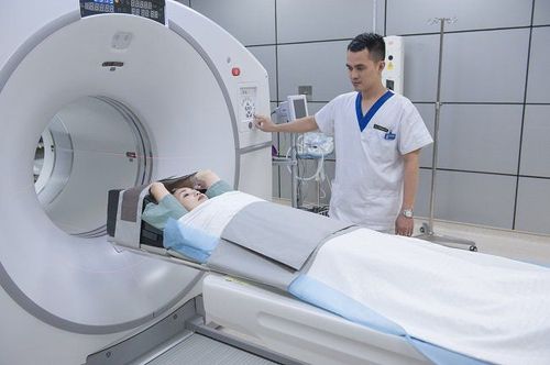
● Prepare patient: review patient records to ensure correct administrative information such as full name, age, occupation and hometown. The patient should be explained and advised on the next steps to be taken. The patient should be instructed to lie in a supine position, with both arms above the head and exposing the chest area to be examined.
Take the shot: set the average slice thickness from 5 mm to 10 mm. Observe the patient's thorax from transverse and anteroposterior views with different imaging windows including skeletal, parenchymal, and mediastinal windows. If contrast injection is performed on a chest CT scan, the patient will be prepared with an IV line and the contrast agent will be administered intravenously. Computed tomography of the chest with contrast injection is indicated mainly in lung cancer, pleural cancer and vascular abnormalities.
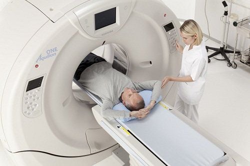
Bệnh nhân được hướng dẫn nằm ngửa khi chụp cắt lớp vi tính lồng ngực
Computed tomography of the chest is a modern imaging method that allows doctors to observe images of the internal structures of the chest to aid in the diagnosis, detection and monitoring of diseases in the chest. chest, heart, lungs and mediastinum.
At Vinmec International General Hospital, the thoracic computed tomography scan is performed by a team of highly qualified and experienced doctors in the field of diagnostic imaging, along with equipment , modern machines for accurate and fast results.
Ths.Bs Nguyen Viet Thu has more than 20 years of experience working in the field of diagnostic imaging, former Secretary of the Department of Radiology in Hanoi, directing the lower level in the field of Diagnostic Imaging. Currently, the doctor is working at the Department of Diagnostic Imaging and Nuclear Medicine - Vinmec Times City International Hospital.
To register for examination and treatment at Vinmec International General Hospital, you can contact the nationwide Vinmec Health System Hotline, or register online HERE