This is an automatically translated article.
The article is professionally consulted by Master, Doctor Le Anh Viet - Radiologist - Department of Diagnostic Imaging and Nuclear Medicine - Vinmec Times City International Hospital.1. Overview of whole body computed tomography
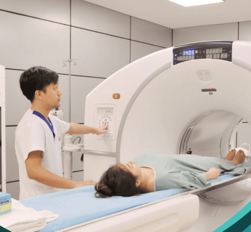
Whole-body computed tomography or full-body CT scan is the name for an imaging method that uses high-intensity X-rays of a helical chamber. Whole-body computed tomography is capable of imaging all vital structures inside the body, including brain computed tomography, abdominal computed tomography, and cage computed tomography. chest.The method of computed tomography appeared in the 1970s, and is now increasingly widely used thanks to its advantages. X-rays are emitted and shined through body tissues in different cross-sections, then converted into images through a processing machine system.
Whole-body computed tomography is a non-invasive technique and is capable of producing 3D images by reconstructing the internal structures of the body based on slices.
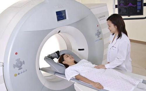
The time to conduct a full-body computerized tomography scan is not too long, on average about 40 minutes to 60 minutes (the time the client is in the scanner is only about 3-5 minutes, the rest is preparation and monitoring time). after shooting). The patient is placed on the bed and taken to the imaging chamber in turn from head to toe. After completing the full-body computed tomography scan, the patient can go home and return to work and normal activities. The doctor will evaluate the resulting images and conduct readings to detect and diagnose the observed imaging abnormalities.
2. What are the highlights of a full-body computed tomography scan?
The advent of computerized tomography technology has brought the Nobel Prize in medicine to two people who discovered it, British physicist Godfrey Hounsfield and doctor Allan Cormack. This event is a testament to the value and role of computed tomography in general in the diagnosis and detection of diseases.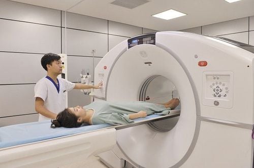
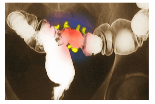
● Capable of detecting and screening cancer at an early stage. Malignant diseases diagnosed at an early stage are very important, helping to increase the effectiveness of treatment and bring the patient's chances of survival.
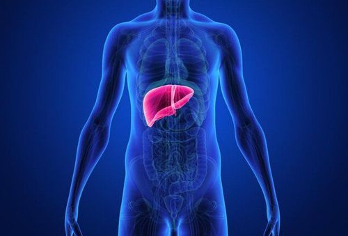
● Whole body computed tomography scan is an opportunity to examine and review the structural and imaging features of all major organs in the body such as brain, heart, lung, liver, spleen, pancreas, and urinary tract. bowel helps detect early stage tumors. Doctors can rely on the images obtained to simultaneously diagnose different diseases in the body and give advice to help prevent complications as well as reduce the chance of metastasis.
● Whole body CT scanning method is an advanced technique but does not require too much from the patient. This is also a safe method and does not require any tools to be inserted inside the subject's body. This contributes to reducing anxiety and pain, helping patients feel more secure during the whole-body CT scan.
However, whole body computed tomography still has its own disadvantages and risks such as:
● The possibility of radiation contamination due to the mechanism of action using high intensity X-rays. Some studies have shown that this can increase the risk of cancers by 10% compared with people without X-ray exposure. contrast if desired to better assess tissue features as well as drug absorption and perfusion characteristics. An allergic reaction to the contrast medium is a risk in this case.
Whole-body computed tomography is not recommended for those at risk, including people with a history of contrast agent allergy, people with chronic kidney failure, women who are suspected of being pregnant or are pregnant. pregnant and young children.

3. Who should get a full-body computed tomography scan?
The main purpose of the whole body computed tomography scan is to screen and diagnose the disease at an early stage, to monitor the general health. However, despite the benefits, there are many risks and disadvantages, and whole-body computed tomography should only be performed in high-risk subjects.The recommended common audience is a group of people older than 50 years old with the ability to detect malignancies, small-sized tumors in organs, spinal diseases... malignancies are more advanced and should be carefully consulted before performing a full-body computed tomography scan.
For people aged 30 to 50 who want to have a full-body computed tomography scan, it should be considered because of the risk of radiation exposure from the computed tomography machine. Instead of whole-body computed tomography, the group of people over 30 years old should only be surveyed in each localized organ area based on clinical symptoms.
Computed tomography is a safe method and has almost no absolute contraindications. Some people who are not recommended to undergo the scan include women who are suspected of being pregnant or are in the first trimester of pregnancy, people with a history of allergy to contrast, and people with chronic kidney disease.
Currently, Vinmec International General Hospital is equipped with a 640-slice CT scanner TSX - 301C manufactured by Toshiba capable of supporting diagnosis on an area up to 16 cm wide, fast speed allows one rotation of the image. The whole heart can be detected, helping to optimally diagnose coronary, vascular and systemic arteries. In particular, this machine can reduce up to 90% of radiation dose, so it can be taken for pregnant women when indicated.
Master Doctor Le Anh Viet has 07 years of experience working in the field of diagnostic imaging, performing well in diagnostic imaging techniques. Currently, the doctor is working at the Department of Diagnostic Imaging and Nuclear Medicine - Vinmec Times City International Hospital.
Please dial HOTLINE for more information or register for an appointment HERE. Download MyVinmec app to make appointments faster and to manage your bookings easily.
