This is an automatically translated article.
The article was professionally consulted by Doctor Head of Department of Medical Examination & Internal Medicine, Vinmec Hai Phong International General Hospital
For patients with coronary artery disease or suspected disease, the doctor may ask the patient to perform some exploratory diagnostic methods to identify the disease. In particular, computed tomography is considered the gold standard method to diagnose coronary artery disease.
1. What is coronary heart disease?
Coronary artery disease includes: coronary atherosclerosis, coronary artery spasm, arteritis in collagen disease and congenital malformations. This condition can be caused by degeneration of the vascular wall or the accumulation of lipid-containing plaques that obstruct blood flow. The disease is common in people with diabetes, high blood pressure, fatty blood, people who are often stressed or smoke too much.
For patients with coronary artery disease, the first symptom is usually angina pectoris. However, many patients do not have chest pain or atypical angina. If coronary artery disease is suspected, the doctor will ask the patient to perform several tests to confirm the disease. Early diagnosis of coronary artery disease plays an important role in the prevention, treatment, and reduction of mortality.

2. Diagnostic methods for coronary artery disease
2.1 Electrocardiogram This is the simplest method to find signs of coronary artery disease. Electrocardiogram can detect myocardial ischemia, myocardial necrosis, coronary artery disease complications such as dilated heart chambers, thickened heart wall or arrhythmia.
Electrocardiogram is a simple, bleeding-free, low-cost, and short-time (sometimes as little as 5 minutes) exploratory method. However, there are quite a few cases where the electrocardiogram is not accurate, such as not detecting coronary disease or identifying the disease in people without disease. Therefore, this method is only intended as an adjunct in detecting coronary artery disease.
2.2 Echocardiography Echocardiography is a method of assessing the movement of the heart walls. In patients with coronary artery disease, the region of myocardium supplied by that coronary branch will not receive enough oxygen, leading to dyskinesia (decreased movement or inactivity) compared with other regions and clearly displayed during ultrasound, helping doctors diagnose the disease accurately.
Ultrasound is a diagnostic method without bleeding, but usually only detects coronary artery disease at a late stage, when the disease has caused cardiac chamber movement disorders.
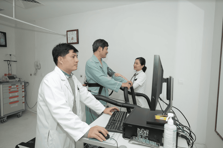
2.3 Stress testing Stress testing is known as the classic way to diagnose coronary artery disease. Normally, at rest, the coronary arteries, even if narrowed, are still able to meet the body's needs. However, with exercise, the body's need for oxygen increases, which will reveal signs of myocardial ischemia. Cardiac ischemia during exercise should be assessed by stress echocardiography, stress electrocardiogram, or stress myocardial scintigraphy. Thereby, the doctor will assess whether the patient is likely to have coronary artery disease and how severe the disease is.
2.4 Exploration and diagnostic imaging The methods of imaging diagnostics such as coronary multi-segment computed tomography, perfusion radiography, cardiac magnetic resonance imaging,... are being widely applied. for early diagnosis of coronary artery disease.
Multi-coronary computed tomography (CT) scan provides anatomical images of the coronary arteries, showing the degree of coronary calcification, coronary artery stenosis, degree of stenosis, the number of coronary artery segments as well as the number of coronary artery segments. other anomalies.

3. Details of the method of computed tomography to diagnose coronary artery disease
Coronary angiography is a procedure done to diagnose the condition of the coronary arteries (the arteries that supply blood to the heart).
3.1 Indication for computed tomography The people who need computed tomography to diagnose coronary artery disease include:
People with risk factors for coronary heart disease such as diabetes, hypertension, hyperlipidemia, addiction heavy smoking, family history of coronary artery disease; People suspected of having coronary artery disease when symptoms of chest pain and ECG, stress electrocardiogram do not clearly identify abnormalities; People who have been treated for coronary artery disease by balloon angioplasty, percutaneous coronary intervention or coronary bypass surgery,... should be monitored after treatment; People need to identify some cardiomyopathies such as enlarged myocardium, abnormality on heart valves. 3.2 Contraindications to computed tomography The cases where computed tomography should not be taken to diagnose coronary artery disease include:
Not cooperating; History of bronchial asthma; Allergy to iodinated contrast medium; Patients with renal failure; Pregnant; People with irregular heartbeat, atrial fibrillation; Person with metallic material in the body. 3.3 How is computed tomography performed? Coronary angiography is done through the groin, wrist, or anterior part of the elbow (mostly the right side). Cardiac catheterization and coronary computed tomography were conducted in the cardiovascular room, intervention with modern angiographic equipment and fluorescent screens.
During the procedure, the doctor will numb the area, insert the catheter into the base of the aorta (the largest artery). The contrast material is then injected into the coronary artery through this catheter. Contrast allows the doctor to see the shape and size of the coronary artery on the fluorescein screen, assess the location and extent of the narrowing of the coronary artery. Percutaneous coronary angiography is a method of exploratory bleeding but is completely painless and very rare with complications.
4. Where is the coronary computed tomography scan?

Vinmec Hai Phong International General Hospital is currently applying computed tomography of coronary arteries and the heart without beta block for patients who need to diagnose coronary heart disease. Choosing to perform the procedure at Vinmec Hai Phong, you will have the most reassuring experience with the following advantages:
Standardized imaging techniques and up to 95% correct diagnosis rate; Paid by health insurance, supported from 61-76%; Equipped with Aquilion One (Vision Edition) 640-slice CT scanner with fast scanning speed, possessing the latest 3D AIDR dose reduction technology, allowing 75% reduction in the dose of rays on patients; The device is capable of synthesizing 320/640 slices in one revolution at a speed of 0.275*s/0.35s; The system uses 160mm wide acquisition Detector with 320 Detector rows, allowing to capture heart, brain... in one rotation; iStation helps to display ECG signals, guide breathing on Gantry; SureExposure 3D continuously adjusts the imaging current during the helical scan in the three spatial axes of X, Y and Z according to the patient's body shape to reduce the dose to the lowest possible; Voice and system patient guidance (Voice link); Boost3DTM allows to reduce the dose to areas with high X-ray absorption such as the shoulder area, helping to acquire images with high accuracy; 3D color image processing (volume reconstruction, surface reconstruction, MPR, curved MPR, MIP, Cine). The team of doctors is comprised of radiologists with a Master's degree, a specialist I with extensive experience, who have worked at first-class hospitals in the region, specifically: Doctor, Doctor Doan Xuan Sinh: has 17 years of experience in diagnostic imaging; Specialist Doctor I Nguyen Dinh Hung: has over 10 years of experience in the field of diagnostic imaging (Ultrasound, CT, MRI); Doctor, Doctor Pham Quoc Thanh: has 13 years of experience in the field of diagnostic imaging, is highly trained in diagnostic imaging domestically as well as internationally.
Please dial HOTLINE for more information or register for an appointment HERE. Download MyVinmec app to make appointments faster and to manage your bookings easily.


