This is an automatically translated article.
Mediastinum is located in the center of the ribcage, contains many important parts of the body. Each organ in the mediastinum has its own disease manifestations.
1. What is the mediastinum?
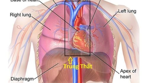
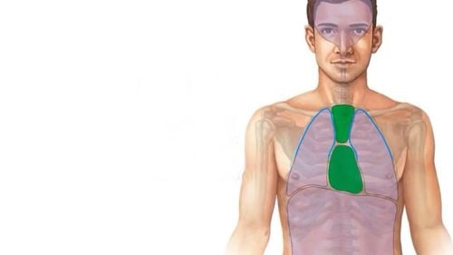
The mediastinum is a cavity located in the thorax, bounded by the base of the neck above, the diaphragm below, the sternum anteriorly, the vertebrae posteriorly, and the parietal pleura bilaterally.For convenience in clinical treatment, the mediastinum is divided into three regions:
Anterior mediastinum Middle mediastinum posterior mediastinum. 1.1 Anterior mediastinum The anterior mediastinum contains the thymus or remains of the thymus, thyroid and parathyroid glands in the thorax, lymphatics and lymph groups in the thorax, fatty tissue, and connective tissue.
1.2 Middle mediastinum The middle mediastinum includes the heart, pericardium, trachea, bronchi, arch of the aorta and great blood vessels, veins and superior venous sinuses, hilum, lymph nodes, nerves diaphragm and X cord.
1.3 Posterior mediastinum The posterior mediastinum includes the esophagus, thoracic duct, lymph nodes, inferior segment of the X nerve, descending aorta, Azygos vein, sympathetic ganglia, and connective tissue.
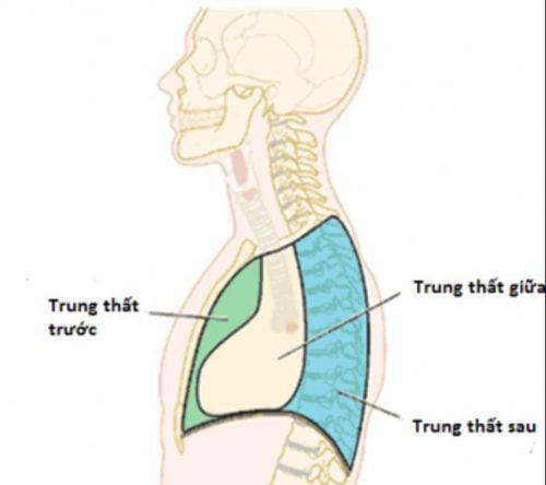
Trung thất được chia làm 3 vùng khác nhau trên giải phẫu
2. Common mediastinal diseases
2.1. Pneumomediastinum There are two types of pneumomediastinum disease: idiopathic pneumomediastinum with identifiable cause and spontaneous pneumomediastinum of unknown cause.Etiological pneumomediastinum are those cases where the cause of the pneumomediastinum is well-known, such as alveolar rupture, tracheobronchial or esophageal sever, trauma, perforation of a hollow viscera, infection or surgery,... Spontaneous mediastinal pneumothorax is the occurrence of mediastinal free air with no known cause. The mechanism of spontaneous pneumomediastinum may occur due to the pressure difference between the alveoli and the pulmonary interstitium leading to alveolar rupture. , dyspnea With spontaneous pneumomediastinum, subcutaneous emphysema may occur. Treatment of mediastinal pneumothorax depends on the underlying cause. Treatment of spontaneous pneumomediastinum is mainly supportive treatment, including measures such as resting, giving the patient oxygen, using prophylactic antibiotics, bronchodilators,...
2.2. Mediastinitis 2.2.1. Acute mediastinitis Acute mediastinitis is an infection of the loose connective tissue in the mediastinum, which surrounds the organs in the mediastinum. This is a very severe, rapidly progressive mediastinal disease that can be fatal in a short time. Acute mediastinitis can be caused by:
Local infection: Due to bacterial infection of the esophagus, tracheal trauma, puncture or scratched mucosa due to endoscopy, fishbone... grow, spread, penetrate the connective tissue, penetrate the mediastinum, cause mediastinal infection. Infections at sites close to the mediastinum spread to such as osteomyelitis of the cervical vertebrae, pleurisy, pneumonia, foci of osteomyelitis of the sternum,...

Viêm phổi do nhiễm khuẩn có thể gây viêm trung thất cấp
Infections from a distance to the mediastinum such as infections from the neck to the mediastinum, infections from the abdomen to the mediastinum,... Acute mediastinitis has symptoms of severe infection such as: high fever, chills, difficulty breathing, pale face, rapid pulse, can be severe pain in the sternum, edema of the base of the neck above the sternum, emphysema under the skin,...
This is a mediastinal disease requiring emergency treatment and intensive resuscitation. Combined poles, for patients using high-dose antibiotics, opening mediastinal drainage, if there is a wound in the esophagus that cannot be sutured, a gastrostomy procedure is performed to nourish the patient.
2.2.2. Chronic mediastinitis Chronic mediastinitis includes two main conditions, mediastinal fibrosis and mediastinal granulomatous inflammation.
Mediastinitis is a disease that causes fibrosis of the connective tissues of the mediastinum, the disease often causes compression of the trachea, blood vessels and nerves, and paralysis of the recurrent laryngeal nerve and the phrenic nerve. Mediastinal granulomatosis: Symptoms of the disease are often poor, diagnosed by chest X-ray film and endoscopic mediastinal lymph node biopsy. 2.3. Mediastinal tumor 2.3.1. What is a mediastinal tumor? Mediastinal tumors are the most common mediastinal pathology, accounting for more than 90% of cases, of which the majority are malignant mediastinal tumors. Mediastinal tumors are common according to certain locations in the mediastinum:
Common tumors in the anterior mediastinum include thymoma, thymic cyst, germ cell tumor, thyroid goiter, parathyroid adenoma, tumors lymphomas, connective tissue tumors. Common tumors in the mediastinum include tracheal tumors, lymphomas, and cancer metastases. Common tumors in the posterior mediastinum include neoplasms, nerve cysts, esophageal tumors, pancreatic pseudocysts, and diaphragmatic hernias.
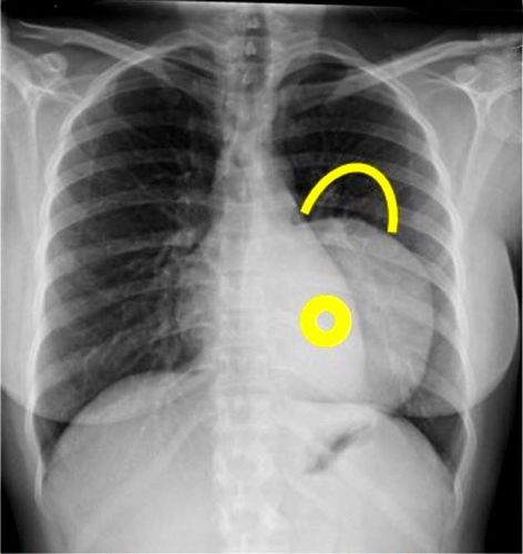
Hình ảnh U trung thất trên kết quả chụp phim Xquang của người bệnh
2.3.2. Symptoms of mediastinal tumor The common symptoms are:
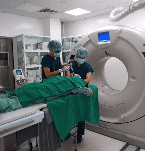
Whole body fatigue, fever, loss of appetite, weight loss,... due to the tumor pressing and causing respiratory tract infection. Cough, difficulty breathing, wheezing or wheezing, sometimes coughing up blood Difficulty swallowing due to tumor pressing on esophagus Chest pain due to tumor pressing on intercostal nerve; hoarseness due to compression of the recurrent nerve; breathing disturbances, high blood pressure, increased salivation due to the tumor pressing on the X nerve; pupillary constriction, eyelid drooping, narrowing of the eyelid slit, flushing on one side of the face, ... due to compression of the cervical sympathetic plexus. When the tumor compresses the superior vena cava causing symptoms of swelling of the face, shoulders, neck, fullness of the supraclavicular pit, cyanosis of the lips, headache, ... compression of the inferior vena cava causes hepatomegaly, ascites, and lower extremity edema; Single vein compression causes collateral circulation in the chest wall. If the mediastinal tumor compresses the thoracic duct, chylous pleural effusion, chylous ascites, and lower extremity edema may be seen gradually spreading to the upper extremities. If the tumor compresses and enlarges, it can cause edema and swelling in the chest wall and breastbone adjacent to the tumor. 2.3.3. Treatment of mediastinal tumor Diagnosis of mediastinal tumor by clinical examination, combined with subclinical techniques such as chest X-ray, computed tomography (CT), magnetic resonance imaging (MRI), endoscopy and examination tumor biopsy,...
All mediastinal tumors have been confirmed, if there are no contraindications, they should be surgically removed early. Surgery is both a treatment method and helps diagnose advanced stages, histopathology, thereby helping to determine additional treatment plans after surgery such as chemotherapy, radiation therapy,...
Surgical methods Surgery for some common mediastinal tumors:
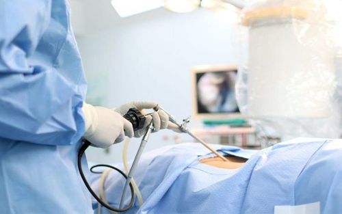
Neuromas: Usually endotracheal anesthesia and surgery through the lower posterior thoracotomy due to neuromas are usually in the posterior mediastinum. If you have Schwann's capsule, you can use thoracoscopic surgery. In the case of malignant neuroma that strongly invades surrounding organs, sometimes surgery only removes part of the tumor.
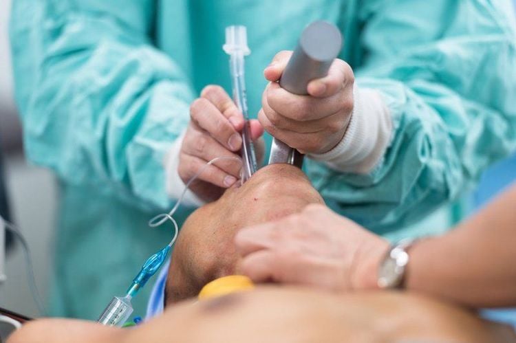
Người bệnh được gây mê nội khí quản trước khi phẫu thuật u trung thất
Monster tumor: Usually in the anterior mediastinum, so the opening along the middle of the sternum is the most appropriate incision. Difficulty in surgery for mediastinal teratoma is that the tumor often sticks to surrounding organs, so the surgical process can easily cause damage to that organ. Thymus tumor: If thymic tumor has symptoms of severe myasthenia gravis, medical treatment is required before surgery. The most favorable incision is to open the sternum along the midline of the upper 2⁄3 or the entire sternum. Any questions that need to be answered by a specialist doctor as well as if you want to be examined and treated at Vinmec International General Hospital, you can contact Vinmec Health System nationwide or register online. online HERE.


