This is an automatically translated article.
Deep vein thrombosis is a fairly common disease, capable of being blocked by a blood clot. This disease can occur in veins throughout the body but is most likely to be in the lower extremities, specifically the thighs or calves. Color Doppler ultrasonography is the primary method for the diagnosis of deep vein thrombosis.
1. What is deep vein thrombosis of the lower extremities?
The venous system consists of 3 types, namely superficial veins, deep veins and perforating veins. These 3 types of veins function to carry blood after exchanging oxygen from the organs back to the heart. Veins have one-way valves that move blood in a certain direction.
Deep vein thrombosis of the lower extremities is a condition in which a blood clot forms on the inside of a vein in the calf or thigh. The main cause of this disease is the decrease in venous return, thereby causing endothelial dysfunction and hypercoagulability. There are three factors that contribute to the formation of venous thrombosis, namely circulatory stasis, venous endothelium damage, and hypercoagulability.
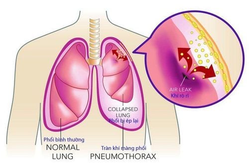
This disease, if not treated promptly, will cause a lot of complications such as skin ulcers at the site of blood clots, leg pain, leg edema or, most dangerously, pulmonary embolism. Because when there is a thrombus, the vein that carries blood flows to the right atrium and then to the right ventricle. At this time, the right ventricle pushes the clot to the lungs, but the blood vessels in the lungs are small, so it will be blocked by the blood clot.
2. Signs of deep vein thrombosis of the lower extremities
Deep vein thrombosis of the lower extremities often has no obvious symptoms, only causing pain and swelling when the condition is too severe. But it is still possible to detect the disease by some easy-to-see signs such as:
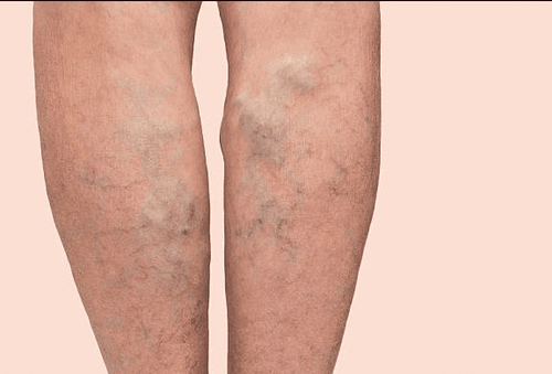
Swollen feet, heavy legs, can see a clear difference between the legs. Areas of skin affected by deep vein thrombosis tend to turn blue-black. Painful feeling when walking. Fevers often occur without a cause. Feeling hot feet. The veins in the legs are clearly visible. More serious complications such as difficulty breathing, coughing a lot, coughing up blood, chest pain, etc. Pain along the veins. Swollen feet, pressing on the legs will concave.

Siêu âm doppler màu là phương pháp chủ yếu để chẩn đoán huyết khối tĩnh mạch sâu.
3. The main cause of deep vein thrombosis of the lower extremities
There are direct causes of lower extremity deep vein thrombosis and predisposing factors that increase the risk of the disease.
Direct causes of deep vein thrombosis in the lower extremities:
People with underlying malignancies such as cancers of the lung, ovary, pancreas, urinary tract, stomach... accompanied by hypercoagulability. Fracture of femur, fracture of vertebrae,... Venous insufficiency. Congenital blood clotting disorder. Long periods of immobilization lead to circulatory stagnation due to paralysis, muscle weakness or castration of the lower extremities. Surgery on chest, abdomen, knee, hip... orthopedic due to fracture. People with malignancy. Risk factors for deep vein thrombosis of the lower extremities:
The older you are, the more likely you are to experience deep vein thrombosis of the lower extremities. Obstruction of blood flow to the heart, increased blood clotting causes circulatory stagnation during pregnancy. Long-term use of oral contraceptives or estrogen therapy. People with a history of myocardial infarction, stroke, heart failure. Fat. Lazy exercise. Smoke a lot of cigarettes.
4. Diagnosis of deep vein thrombosis of lower extremities by color doppler ultrasound
There are many ways to diagnose deep vein thrombosis of the lower extremities such as B-Mode ultrasound, D-dimer test,... In which, the most used method is color doppler ultrasound. Because this is the most modern method today, with high accuracy and color images, it is easier to observe.
Color doppler ultrasound is divided into two types as follows:
Continuous color doppler ultrasound: It can be understood as an ultrasound method consisting of two crystals in the same transducer with two completely different functions. One crystal is responsible for emitting sound waves, the other end is used to receive sound waves. As a result, continuous Doppler can be checked very quickly. Reverse pulsed color doppler ultrasound: This technique has only one crystal at the tip, so it can both pick up sound waves and emit sound waves. In particular, pulsed color doppler ultrasound can adjust the size of the area to be examined. The disadvantage of this method is that it is not possible to capture the entire sound wave, but only the sound wave at the test site. Due to its ability to check the movement of the veins, color doppler ultrasound is therefore particularly effective in diagnosing deep vein thrombosis of the lower extremities. Color Doppler helps to define the boundaries of the thrombus and confirm the diagnosis of non-adherent thrombus. This method can also determine whether a vein is completely occluded or semi-occlusive based on the received acoustic wave signal, the anteroposterior variation of the running speed, and the coloration at the site of the occlusion. If the occlusion is semi-occluded, the color flow will move when the muscle is squeezed, and when the color flow does not change, it means it is completely blocked.
Therefore, when the diagnosis by color Doppler ultrasound method gives positive results, it is possible to confirm the diagnosis of the patient with deep vein thrombosis of the lower extremities. In the case of negative ultrasound results, additional D-dimers or Doppler ultrasound will be done within a week.
Deep vein thrombosis is a dangerous disease that requires early and careful examination to avoid complications of pulmonary embolism. Therefore, when having symptoms of the disease or suspecting the disease, customers should immediately make an appointment with the doctors of Vinmec International General Hospital for timely examination and appropriate diagnosis and treatment.
Please dial HOTLINE for more information or register for an appointment HERE. Download MyVinmec app to make appointments faster and to manage your bookings easily.