This is an automatically translated article.
The article is professionally consulted by Master, Doctor Lam Thi Kim Chi - Radiographer - Department of Diagnostic Imaging - Vinmec Danang International General Hospital.Chest X-ray is a modern imaging technique to examine and evaluate the entire internal structure of the lungs, helping to detect lesions and pathologies in the lungs. Chest radiography includes many techniques such as projection, straight chest X-ray, tilting chest X-ray, computed tomography, contrast pump bronchogram, high voltage ....
1. What is a chest X-ray?
Chest X-ray is a method of using an X-ray machine to observe and reconstruct the internal structure of the lungs, thereby helping to detect lesions and pathologies in the lungs.X-ray machine will emit short-wavelength X-ray radiation that passes through soft tissue cells and body fluids to record images of the lungs on an x-ray.
2. What methods of chest X-ray are there?
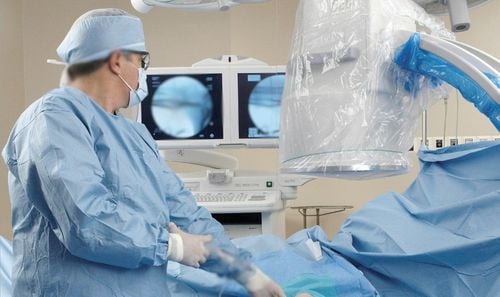
Chest radiography includes many techniques such as projection, straight chest x-ray, tilting chest x-ray, computed tomography, contrast pump bronchogram, high voltage....2.1 Straight chest X-ray An upright chest x-ray is a method of taking a patient in a standing position and the direction of the scan is from back to front. In case the patient cannot stand, the scan should be taken in the semi-reclining position (also known as the Fowler position) and the patient should not be taken in the lying position because of limitations in diagnosing some lung lesions such as: effusion or pneumothorax.
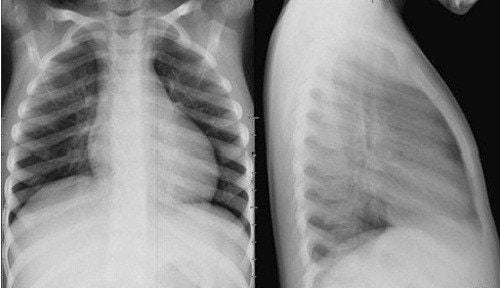
Hình ảnh chụp X-Quang phổi thẳng
Capture both lung fields. Two shoulder blades separated with bilateral lung fields. The image shows 5 vertebrae from D1-D5 and the 6th intercostal space on the diaphragm. In order to obtain clear images, the patient should be instructed to take a deep breath and hold their breath during bronchoscopy.
2.2 Tilt X-ray - Tilt chest X-ray is a method of taking the patient in a standing position and the direction of the scan is from left to right or vice versa, depending on which side the suspected lesion is located.
Tilt chest x-ray also allows analysis of lung parenchyma and helps clarify cases of suspected mass that cannot be identified by straight chest x-ray. The purpose of a lateral chest x-ray is to partition the mediastinum and identify the lesion in a lobe or lobe of the lung.
An x-ray of the lung should be taken to ensure that the following technical standards are met:
Take a clear picture of the two diaphragms, if taken on a right side, the two right diaphragms are parallel, if taken on a left side, the two diaphragms intersect at 1⁄ 3 later. Photograph of the contralateral flank arch 1.5 cm from the CSL. The image clearly shows the space before and behind the heart. 2.3. Computed tomography was performed when conventional radiographs still did not clearly identify the lesion. This method is very valuable in diagnosing lung, mediastinal and bronchial tumors. High-resolution computed tomography is considered the gold standard in the diagnosis of bronchiectasis.
2.4. Other methods Other methods such as irradiation, bronchoscopy with contrast pump, high voltage imaging... are rarely used nowadays.
Trắc nghiệm: Làm thế nào để có một lá phổi khỏe mạnh?
Để nhận biết phổi của bạn có thật sự khỏe mạnh hay không và làm cách nào để có một lá phổi khỏe mạnh, bạn có thể thực hiện bài trắc nghiệm sau đây.3. What is the purpose of chest X-ray?

Các phương pháp chụp X quang phổi và những điều cần biết
Monitor and check the current condition, internal structure of the lung, help detect early abnormalities or damage in the lungs, thereby providing treatment appropriate treatment. Check for pulmonary vascular abnormalities such as pulmonary arteries and veins. Monitoring the recovery in patients after thoracic surgery.
4. In what cases need a chest X-ray?
Lung X-ray should be taken in the following cases:Suspected lung cancer: Some typical symptoms of lung cancer such as frequent chest pain (in a certain location), dry cough, coughing up blood and sputum , continuous and prolonged cough, loss of appetite, loss of appetite, weight loss, fatigue; In the advanced stage, lung cancer patients may also lose their voice, hoarseness, bones and joints in the fingertips, feet, shoulders, etc. When these symptoms appear, the patient should go to the facilities. medical examination and diagnosis by taking X-rays of the lungs and performing general tests. Chest trauma: Cases of chest trauma with suspected lung injury should be taken with chest X-ray for diagnosis and early detection of injuries such as pulmonary contusion, pleural effusion, ... Regular physical examination Periodic health checkup, including chest X-ray is very necessary and useful because this method will help check and detect lung diseases early when the disease has no symptoms.

Trường hợp bệnh nhân thường xuyên tức ngực, ho ra máu nên chụp X-Quang phổi
5. Things to keep in mind when taking a chest X-ray
To obtain clear images and accurate results when taking a chest X-ray, the patient should note the following information:Remove jewelry, change hospital clothes to limit the influence of metal. during shooting. Coordinate and follow the instructions of the doctor and the imaging technician so that the chest X-ray can be taken quickly and smoothly. Currently, chest X-ray gives quick results. Therefore, after taking the scan, patients only need to wait for a short time to receive the results. Through imaging, the doctor will show the current condition of the lungs. For patients who have undergone chest X-ray many times to check and monitor, it is important to bring all the results of the scans with you to the follow-up examination so that the doctor has a basis for making the best diagnosis. Currently, chest X-ray is an imaging technique widely applied in the diagnosis and early detection of lung lesions and pathologies.
Chest X-ray is an imaging method used routinely at Vinmec International General Hospital to examine and diagnose many diseases, including lung diseases. The process of upright chest X-ray, inclined chest X-ray is performed methodically and in accordance with the standard procedure by a team of highly skilled doctors and nurses, modern machinery system, thus giving results. accuracy, contributing significantly to the identification of the disease and the stage of the disease.
MSc Doctor Lam Thi Kim Chi graduated with a Master's degree in Radiology - Hue University of Medicine and Pharmacy and has over 6 years of experience as a radiologist. Dr. Chi used to work at Da Nang Obstetrics and Gynecology Hospital before working as a radiologist at Vinmec Danang International General Hospital as it is today.
Please dial HOTLINE for more information or register for an appointment HERE. Download MyVinmec app to make appointments faster and to manage your bookings easily.