This is an automatically translated article.
This article is professionally consulted by Master, Resident Doctor Nguyen Van Anh - Radiologist - Department of Diagnostic Imaging and Nuclear Medicine - Vinmec Times City International General Hospital.Ultrasound is one of the imaging methods useful in helping to detect pancreatic cancer. Some studies show that ultrasound can be up to 94% sensitive in diagnosing pancreatic cancer. However, the diagnostic effectiveness of ultrasound is highly dependent on the experience of the technician, radiographer, and the patient's condition.
1. Pancreatic cancer
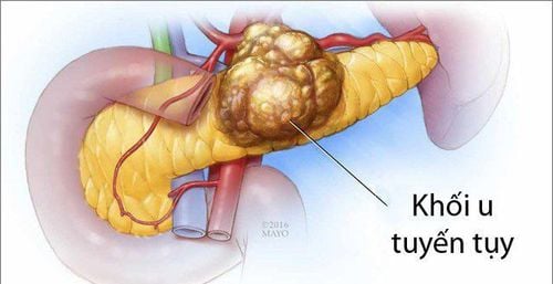
Pancreatic cancer is a type of cancer that occurs when the cells of the pancreas grow abnormally. In the majority of patients, malignancies usually originate in the exocrine pancreas. Tumors originating from the endocrine pancreas are usually benign.How to detect pancreatic cancer? In the early stages of the disease, there are usually no symptoms. By the time symptoms appear, the tumors have grown large and metastasized to other organs.
The symptoms of pancreatic cancer are easily confused with other diseases, especially those of the digestive tract. Therefore, we should not be subjective, we need to see a doctor when one of the following unusual symptoms appears:
● Jaundice: The first symptom, appearing in most cases of pancreatic cancer caused by accumulation of bilirubin – a substance found in bile that the liver secretes to break down fats. Tumor forming in the pancreas grows and compresses the bile duct, causing bilirubin congestion causing jaundice. In addition, when pancreatic cancer has metastasized, one of the earliest affected organs is the liver, leading to the prominent symptoms of jaundice and yellow eyes in the patient.
● Dark urine, pale stools and fat droplets in the stool. In addition to jaundice, these are also symptoms that appear when there is a blockage of bilirubin.
● Abdominal or back pain: When the tumor grows, compresses the organs and nerves around the pancreas, causing the patient to feel pain in the abdomen or back
● Anorexia, weight loss: Unexplained weight loss common in people with pancreatic cancer besides loss of appetite.
● Nausea and vomiting: If the cancer cells multiply, creating a large mass that puts pressure on the stomach, they will cause food to stagnate there, causing nausea and vomiting, especially after eating. .

● Blood clots in large veins, usually in the legs, are called deep vein thrombosis. Diabetes: Sometimes tumors in pancreatic cancer destroy the cells that produce insulin - a substance that helps regulate blood sugar levels, causing diabetes.
There are many ways to diagnose a person with pancreatic cancer, including:
+ Physical exam and medical history: An enlarged liver and gallbladder can be detected when the doctor examines In addition, other risk factors are exploited to find out the cause of pancreatic cancer such as: smoking, drinking a lot of alcohol or family history of cancer.... If there is any abnormality In the test results, the patient may be assigned to do some other tests to be able to make an accurate diagnosis. This test aims to examine abnormal tissues and organs, specifically the pancreas, to determine whether the tumor has metastasized or not, and to provide a prognosis for the disease.
+ Computed tomography (CT) is the optimal method used to diagnose pancreatic cancer because of its ability to provide clear images. In addition, CT can also help determine whether surgery is appropriate in this case.
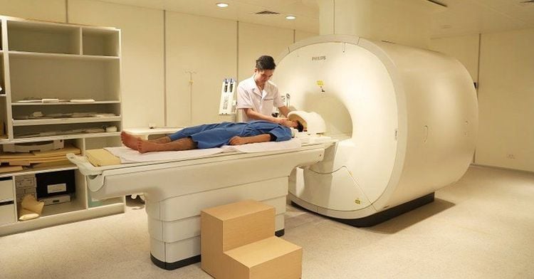
+ Magnetic resonance imaging (MRI): in addition to CT, MRI is also an effective method to help diagnose pancreatic tumors, especially valuable in surveying secondary lesions in the liver and abdomen.

● Endoscopic cholangiopancreatography: Images obtained From this technique helps doctors determine if the bile duct is blocked by tumors or not?
Angiography: A small amount of contrast agent is injected into the artery to outline the structure and function of the blood vessels. This technique helps locate tumors in case they grow and press on surrounding blood vessels. In addition, it also helps identify the presence of abnormal blood vessels that carry blood to nourish cancer cells.
● Blood test: Some abnormal substances can be found in the blood of cancer patients. Finding these substances is the basis for cancer diagnosis.
● Biopsy: A small portion of a suspected cancerous tumor is taken and viewed under a microscope to determine if there are any cellular abnormalities in that area?
2. Ultrasound diagnosis of pancreatic cancer
Ultrasound is one of the techniques used to support the diagnosis of pancreatic cancer, in which the two most commonly used types of ultrasound are:● Abdominal ultrasound: If the patient has symptoms. Abnormalities in the back or abdomen, the doctor may order an ultrasound first because this technique is easy to perform and does not pose any danger to the patient. If the ultrasound results show many signs of pancreatic cancer, the appointment of computed tomography will be considered to help make an accurate diagnosis of the disease.
● Endoscopic ultrasound: Endoscopic ultrasound can provide more accurate results than abdominal ultrasound and is very valuable in the diagnosis of pancreatic cancer. This technique uses a small probe attached to a thin and flexible endoscope, which is slowly inserted into the digestive tract to view tumors or assist technicians in taking biopsies of cancer cells. letters . However, for tumors located far away (the body and tail of the pancreas), endoscopic ultrasound revealed its limitations.

Endoscopic ultrasound is a great step forward in today's gastrointestinal endoscopy. This modern technique is supplementing the limitations of other subclinical diagnostic methods, thereby improving the effectiveness of more accurate and timely disease treatment. Vinmec International General Hospital is a high-quality medical facility in Vietnam with a team of highly qualified medical professionals, well-trained, domestic and foreign, and experienced.
At Vinmec, there are all modern diagnostic facilities such as: PET/CT, SPECT/CT, MRI, blood marrow test, histopathology, immunohistochemistry test, gene test, biological test molecules, as well as a full range of targeted drugs, the most advanced immunotherapy drugs in cancer treatment. Multimodal cancer treatment from surgery, radiation therapy, chemotherapy, hematopoietic stem cell transplantation, targeted therapy, immunotherapy in cancer treatment, new treatments such as autoimmunotherapy body, calorific value,...
Please dial HOTLINE for more information or register for an appointment HERE. Download MyVinmec app to make appointments faster and to manage your bookings easily.
Article referenced source: Cancer.org


