This is an automatically translated article.
Post by Master, Doctor Mai Vien Phuong - Gastrointestinal Endoscopy - Department of Medical Examination & Internal Medicine - Vinmec Central Park International General Hospital.
The appearance and growth of pancreatic cancer is complex and can change the patient's condition. In this regard, the application of artificial intelligence can be applied to diagnose and obtain objective data processing results. However, the application of artificial intelligence in health is not independent and most can only be used as an auxiliary tool.
1. The role of artificial intelligence in pancreatic cancer diagnosis
“Deep Learning” algorithm refers to an ANN image algorithm with many hidden layers. Using this technique, cystic tumor markers, amylase, cytology and other information are entered and then combined with the two data, the output layer indicates whether the pancreatic cystic lesion is benign or malignant. Some researchers have also proposed a framework of supervised classification techniques to extend the diagnosis of pancreatic cancer, provided that the expression profile of a single cell can be imported to reveal its identity.
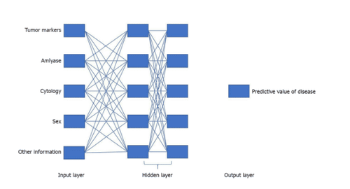
Specific input projects at the beginning of modeling are required for machine learning and neural network analysis. However, for researchers who have not performed data analysis, it is not clear what raw data is and is not needed. Useless data simply increases the workload and can also become the specificity and sensitivity of the model. Meanwhile, model editing also poses a significant problem. While AI can save time, the threshold and workload in setting up AI programs is taboo for non-professionals who lack a background in math and programming. .
The appearance and growth of pancreatic cancer is complex and can change the patient's condition. In this regard, the application of artificial intelligence can be applied to diagnose and obtain objective data processing results. However, the application of artificial intelligence in health is not independent and most can only be used as an auxiliary tool. But with continuous development and improvement, artificial intelligence may eventually have a more universal application.
2. The application system of artificial intelligence must have the combination of experts from many fields
The relationship between the application of artificial intelligence and imaging involves knowledge from various fields such as pathology, radiology, oncology and computer science. Thus, a smarter artificial intelligence system can be built through the combined work of experts from many fields. The artificial intelligence image acquisition discussed is based on segmenting the pancreas from the image. The traditional segmentation method is a top-down simulation fitting method, based on large amounts of map input and fixed pancreatic label fusion. However, there is also a bottom-up pancreatic segmentation method to subdivide the composite image area into pancreatic and non-pancreatic regions. Segmentation based on visual features of images can improve the accuracy of pancreatic segmentation. It is reported that the bottom-up pancreatic segmentation method has been optimized. With the improvement of the deep convolutional neural network, this method can handle the complex appearance of pancreas in CT imaging.
Based on the above, we can see that the application of artificial intelligence in pancreatic cancer diagnosis has made significant strides and is constantly being improved.
3. Application of artificial intelligence in histopathological diagnosis of pancreatic cancer
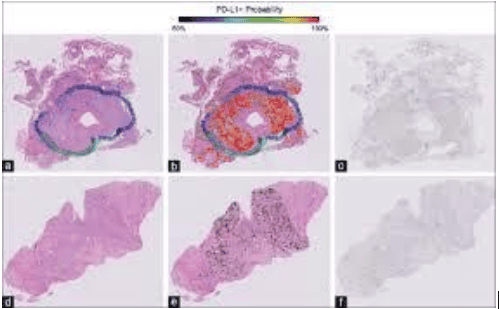
Pathologists need to identify diseased tissues in different tissue sections, which is a time- and labor-intensive process. Even experienced professionals can be at risk due to subjective judgment. As well as the application of artificial intelligence in diagnostic imaging, this method is also important in the field of pathology, where tissue sections are digitized by computer.
First, the artificial intelligence system divides the lumen and nucleus from tissue fragments as well as extracts vector features from the epithelial nucleus tenfold. Different cells have different feature vectors. An epithelial multiplication algorithm is used to identify epithelial nuclei. The morphological features of the disease can then be extracted. Finally, an artificial intelligence classifier is used for classification.
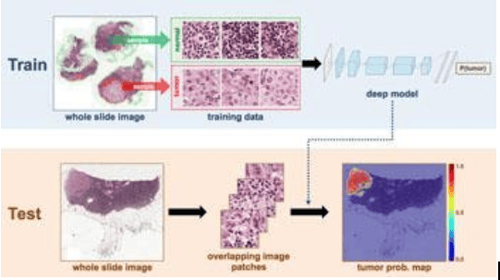
Based on the automatic learning framework, cells can be segmented more precisely by combining bottom-up and top-down information. After the patient's tissue samples were collected, tissue photographs were collected uniformly. The agglomeration neural network model of the deep agglomeration neural network was used to generate a probabilistic map of the tissue nuclear distribution. Then, the repeated region merge method is used to initialize the shape of the probability graph. Next, combining a sparse conformation model with stable selection and a local repulsive strain model, a new segmentation algorithm is proposed to split a single nucleus.
4. The advantage of matching with different histopathological staining images
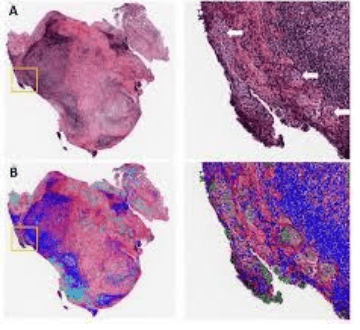
Due to the deep neural network feature learning and high-level conformational features prior to modeling, this proposed method is universal enough and can be applied to staining patterns different histopathology. This model is not only less susceptible to overlap of pathological tissues and cells, but also relatively sensitive to image noise and uneven intensity. Various tissue staining datasets are tested, which can identify and label the nuclear focal region.
5. Neural networks play a role in image analysis
In addition to the artificial intelligence classifier, the neural network also plays an important role in image analysis to determine whether the pathology is benign or malignant. After collecting a certain amount of pathologic fine-needle aspiration of the pancreatic tumor, a pathological image is captured for pre-processing (image grayscale and noise reduction). Then the K-means clustering algorithm is used to extract the highest value of the pixels until all are equal. The portion of the image that needs to be defined is segmented so that the tissue can acquire basic nuclear features, which are used to evaluate cell morphological features. These features are fed into the artificial intelligence multilayer perceptron (a linear nonlinear neural network) as input vectors, and the decisions made by this perceptron are sent to the second layer perceptron using evaluation. image price.
6. The diagnostic accuracy of benign and malignant lesions can be evaluated by statistical methods
Because there are certain confirmed cases, the diagnostic accuracy of benign and malignant lesions can be evaluated using statistical methods (logistic regression, multiple regression, area under curve and R-squared). Unlike imaging, pathology pays more attention to accuracy. Therefore, the application of artificial intelligence has a lot of opportunities to improve the accuracy of adjunctive diagnostics, which will certainly take a long time to develop.
Currently, Vinmec International General Hospital has been implementing cancer screening packages. At Vinmec, there are fully modern diagnostic facilities such as: PET/CT, SPECT/CT, MRI..., blood marrow test, histopathology, immunohistochemistry test, gene test, lab test molecular biology, as well as a full range of targeted drugs, the most advanced immunotherapy drugs in cancer treatment. Multimodal cancer treatment from surgery, radiation therapy, chemotherapy, hematopoietic stem cell transplantation, targeted therapy, immunotherapy in cancer treatment, new treatments such as autoimmunotherapy body, heat therapy...
Thanks to modern facilities, a team of qualified doctors and medical staff, perfect medical services have brought confidence, health and good quality of life to patients. medical examination and treatment at Vinmec.
Please dial HOTLINE for more information or register for an appointment HERE. Download MyVinmec app to make appointments faster and to manage your bookings easily.
References:
1. Adamska A, Domenichini A, Falasca M. Pancreatic carcinoma: Current and developing therapies. Int J Mol Sci . Year 2017; 18 : 1338. [ PubMed ] [ DOI ]
2. Halbrook CJ, Lyssiotis CA. Using metabolism to improve diagnosis and treatment of pancreatic cancer. Cancer cell . Year 2017; 31 : 5-19. [ PubMed ] [ DOI ]
3. Ngiam KY, Khor IW. Big data and machine learning algorithms for healthcare delivery. Lancet Oncol . Year 2019; 20 : e2 62-e273. [ PubMed ] [ DOI ]


