This is an automatically translated article.
Bronchoscopy with fluorescent light can detect carcinoma in situ, dysplastic lesions that are difficult to identify with white light bronchoscopy alone.
1. What is bronchoscopy with fluorescent light?
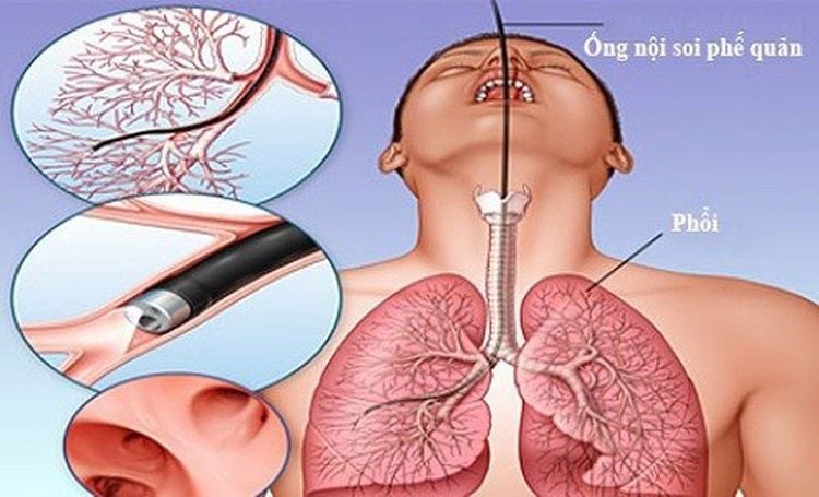
Currently, bronchoscopy has an important role in locating bronchial lesions and biopsies to identify pathology. This method is based on the principle that dysplastic and malignant tissues reduce the autofluorescence signal compared to normal tissues.
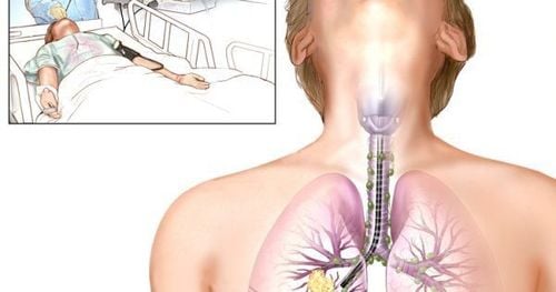
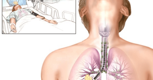
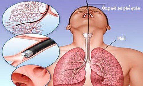
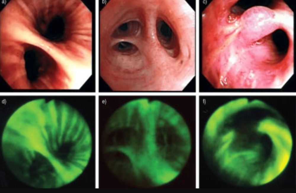
Bronchoscopy with fluorescent light is an endoscopic method with a flexible bronchoscope that uses blue light to observe in order to detect premalignant and malignant lesions that by conventional bronchoscopy. It is usually not detectable based on the principle of different light capture and autoluminescence of tumor tissue and healthy tissue.
The filtered image is processed to put on the screen, the normal organization will be blue and the abnormal will be red. Bronchoscopy with fluorescent light is used to detect dysplastic lesions, tumors in situ, or cancerous invasions that cannot be detected by conventional bronchoscopy.
However, this method has certain limitations, specifically bleeding or inflammatory lesions will give a reddish brown color, so the diagnosis is confirmed based on biopsy results.
Phương pháp nội soi phế quản giúp chẩn đoán và xác định tổn thương
2. Indications and contraindications for bronchoscopy with fluorescent light
Bronchoscopy with fluorescent light is contraindicated in the following cases:
There is a history of allergy to anesthetics, anesthetics. Coagulation disorders Moderate to severe respiratory failure. Heart attack ; severe heart failure.
Người bệnh mắc rối loạn đông cầm máu không nên nội soi
Bronchoscopy with fluorescent light is indicated in the following cases:
There is a previous image of dysplasia; Lung cancer staging. Early detection of bronchial cancer in high-risk subjects such as smokers, waterpipes, etc. positive cell tests. Detecting metastatic lesions or recurrent lesions in the endotracheal lumen for patients who have undergone surgery and chemotherapy for bronchial cancer. There is a suspicion of bronchial cancer in those who cough up blood with no visible lesions on routine bronchoscopy.
3. Steps of bronchoscopy with fluorescent light
Steps of bronchoscopy with fluorescent light are performed as follows:
Step 1: Prepare equipment including: flexible bronchoscope; signal converter system; xenon and white light sources; biopsy pliers: 01 pcs; vaccum; anesthetics, anesthetics, shock absorbers and some other specialized equipment. Step 2: Conduct tests for the patient before the procedure. Step 3: The surgeon numbs the larynx to the patient, then conducts bronchoscopy according to the flexible bronchoscopy procedure. Note: Avoid scratching, congesting or bleeding the bronchial mucosa. Step 4. After placing the endoscope, the procedure operator presses the light system switch button on the endoscope to detect lesions. If lesions are detected, biopsies and cleaning are performed. Step 5: Remove the fluid and blood generated by the procedure and remove the bronchoscope from the patient. Step 6: Instruct the patient to fast for 2 hours after the endoscopy.
Hình ảnh nội soi phế quản với ánh sáng huỳnh quang
Vinmec International General Hospital is one of the hospitals that not only ensures professional quality with a team of leading medical doctors, modern equipment and technology, but also stands out for its examination and consultation services. comprehensive and professional medical consultation and treatment; civilized, polite, safe and sterile medical examination and treatment space.
Customers can directly go to Vinmec Health system nationwide to visit or contact the hotline here for support.
MORE:
Role of bronchoscopy in the diagnosis of respiratory pathology Process and possible risks during bronchoscopy Bronchoscopy in children: Things to note


