This is an automatically translated article.
The article was professionally consulted by Specialist Doctor I Tran Cong Trinh - Radiologist - Radiology Department - Vinmec Central Park International General Hospital. The doctor has many years of experience in the field of diagnostic imaging.Currently, brain biopsies can be performed via open biopsies or localized biopsies. Open biopsies can be easily performed for superficial lesions of the cortical surface. However, for small lesions, especially deep in the brain parenchyma, open biopsy is difficult to obtain tissue samples accurately.
1. Overview
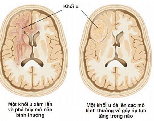
Today, along with the development of imaging tools, especially computed tomography and magnetic resonance imaging, the number of patients with brain lesions is increasing, the need for treatment is increasing. high. The prognosis and selection of appropriate treatment depends on the nature of the lesion. The nature of the lesion is the pathological result from the specimen taken from the pathological mass, which is considered the "gold standard" of the disease. This can be done through open surgery, removing the pathological mass.Surgery is the fastest and most effective method of removing lesions. However, for lesions located deep in the brain parenchyma or in functional brain regions, surgery can cause damage to healthy brain tissue, endangering the patient's life or leaving severe sequelae. more severe. In these cases, taking tissue samples through biopsy plays a very important role.
However, for small lesions, especially deep in the brain parenchyma, open biopsies are difficult to obtain accurate tissue samples. Along with the development of imaging techniques, many positioning systems have been born that make it possible for biopsies to accurately obtain tissue samples, especially helping to minimize damage to healthy brain parenchyma. Biopsies guided by localization systems are called localized biopsies.
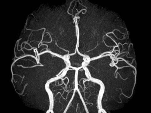
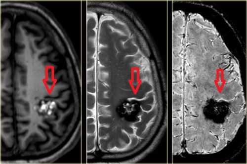
Chụp cộng hưởng từ mạch máu giúp cho việc lập kế hoạch sinh thiết chính xác hơn
2. What is a brain biopsy?
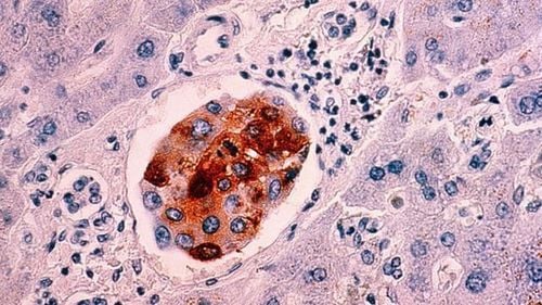
To put it simply, a brain tumor biopsy is taking a small portion of a tumor in the brain for testing. When biopsies, doctors use under the guidance of the navigation system or the nervous system to guide. Specifically, the doctor made a 2cm incision and drilled a small hole in the skull. A fine needle is then inserted into the tumor to take a sample. Pieces of brain tumors are processed through a process and studied under a microscope. After observing and analyzing, the doctor can conclude whether the tumor is benign or malignant. The surgeon can do the biopsy in 3 ways:Needle biopsy: Make a small incision in the scalp, drill a small hole through the skull bone, and then use a needle to aspirate some brain tissue with the tumor. Computerized tomography or MRI guided biopsy. Simultaneous biopsy with treatment : The doctor takes a little tissue and sends it for cytology after removing the brain tumor.
3. Indications for brain biopsy

Sinh thiết não định vị không khung được chỉ định với bệnh nhân u não ác tính
The lesion does not appear to be "stunned" or there is no indication for open surgery such as: multifocal metastatic tumor or malignant brain tumor. Injuries to the deep, functional cortex or deep gray nucleus (basal ganglia, thalamus ..) are locations when surgery will bring high rates of death and disability. Invasive lesions: no clear boundaries, easy to get mixed into healthy brain tissue. Lesions appearing on imaging or clinical manifestations are suspected of inflammatory causes, demyelinating disease.
4. Steps to perform a brain biopsy via computed tomography
Prepare the patient: The patient has a complete medical record and basic laboratory tests including baseline coagulation, HIV and HbsAg. Explain the biopsy procedure to the patient and family.Prepare equipment: Sterile tools and medicine needed for the procedure.
Steps to perform a brain biopsy:
Step 1: Enter image data and the system locates and program the biopsy. Step 2: Fix the reference frame to the Mayfield price. Step 3: Create a link between the computer and the patient. Step 4: Locate the biopsy point on the skin.
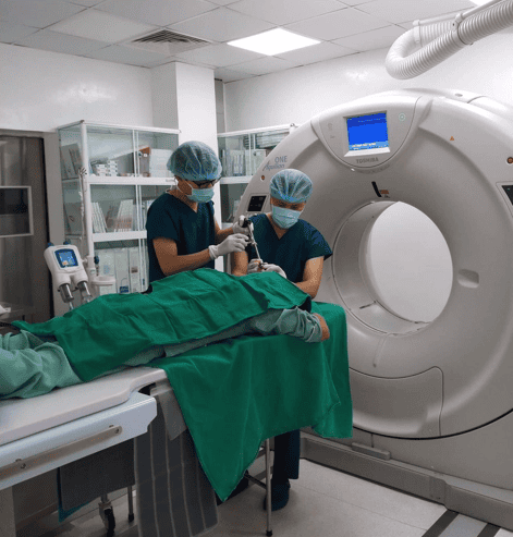
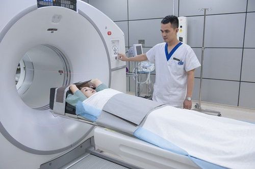
Sinh thiết não qua chụp cắt lớp vi tính
5. Some complications after brain biopsy
Some complications can occur during and after the brain biopsy procedure. However, the complications that occur are all non-serious complications that do not affect the patient's life or function. During follow-up, the patients with these complications all had a stable development after the biopsy, no new focal neurological signs appeared, and had a normal CT image after 2-7 days.Acute complications: Bleeding in the brain tumor, in the brain parenchyma, in the subarachnoid space. Intracranial gas accumulation, sudden loss of consciousness or focal neurological signs.
Late complications: meningitis, osteomyelitis at the biopsy site
Vinmec International General Hospital is a high-quality medical facility in Vietnam with a team of highly qualified, trained medical professionals. Create methodical, specialized domestic and foreign, rich experience.
A system of modern and advanced medical equipment, possessing many of the best machines in the world, helping to detect many difficult and dangerous diseases in a short time, supporting the diagnosis and treatment of doctors the most effective. The hospital space is designed according to 5-star hotel standards, giving patients comfort, friendliness and peace of mind.
Please dial HOTLINE for more information or register for an appointment HERE. Download MyVinmec app to make appointments faster and to manage your bookings easily.


