This is an automatically translated article.
When taking X-ray films, the recognition of normal or abnormal images on the film is very important to help diagnose or guide the diagnosis of bone pathology. The image of bone lesions on x-ray films is very diverse, divided into lesions in terms of contrast, structure or shape of the bone.
1. Normal bone x-ray image
X-ray - bone x-ray is a widely used diagnostic imaging method, using X-rays to shine through the bone and joints to be investigated, from which to obtain images and from observing the acquired image characteristics. which can diagnose the current condition.
The skeletal structure consists of long, bony, flat, and fibula bones. On the X-ray film, the images of bones such as:
Long bones, also known as tubular bones, are composed of bone heads; The bone body has a large radiopaque component consisting of solid bone and a non-contrast canal. The outside is a non-contrast periosteum, so it cannot be seen on film. Flat bones and cubs: The main component is cancellous bone surrounded by a dense but very thin layer of bone, so the contrast is less.

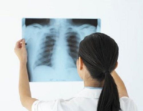
Hình ảnh chụp X-quang xương giúp chẩn đoán hoặc định hướng các bệnh lý của xương
2. Bone lesions on bone x-ray images
2.1 Contrast abnormalities Bone X-ray resistance depends on the concentration of calcium in a unit of bone tissue. They can be altered on film by decreasing contrast or increasing contrast.
Decreased contrast density: This condition is seen when the amount of calcium in the bone is reduced by at least 30%, on x-ray, it is shown as an enhanced image compared to surrounding healthy tissue. This condition is called osteoporosis. This manifestation can be seen in many bones in the body due to osteoporosis or other systemic diseases such as parathyroid disease, rickets, immobilization for a long time... Localized in certain locations can be seen in infections, tumors, trauma... Increased bone contrast: When the ratio of calcium concentration in bone increases, the film shows a blurred image than the surrounding healthy tissue or called a characteristic feature. the bone. Diffuse manifestations are seen in blood diseases, bone metastases, thyroid dysfunction, heavy metal poisoning, bone syphilis, congenital bone diseases. Localized: As a response of bone to injury, pathology with bone neoplasia as regeneration after fracture. It is possible to meet the situation of mixed contrast as in chronic inflammatory disease, tumor... 2.2 Abnormalities in bone structure. This image shows that the bone tissue does not have calcium, so the image is not or hypocontrast. Depending on the extent of bone destruction, we can see images of osteoporosis or bone thinning, bone destruction, bone loss, bone necrosis, and dead bone fragments. If osteoclasts are seen in the bone, suggest metastatic cancer, cancerous tumors of blood origin, or infection. If in the metaphysis, think of primary bone cancer. If the bone ends think of osteoclast tumor, chondroblastoma. If the bone destruction has a clear borderline, there is no reaction around the lesion, the disease will progress slowly, such as Kahler's disease, reticular disease. If the border is discontinuous, it indicates a rapidly progressing lesion such as cancer or acute infection; There are limited cases of opacity with many small defects often due to primary malignancy. Thin bone solids are seen in benign osteosarcomas, thickened bone margins or inflammatory lesions are common. Bone formation Osteogenic reaction: Is the phenomenon of new bone formation starting from the bone fibers, this process causes the bone to thicken and show morphological changes... On the film, images such as dense bone, fibrous appear on the film. bone...
Periosteal reaction: Normally, the periosteum on film is not contrasted, the periosteal reaction is caused by the process of impacting on the periosteum, causing a new bone formation reaction from the inner periosteum. This phenomenon can be caused by tumors or infections. Although it is not specific for any type of bone tumor, it is a feature that distinguishes benign and malignant osteomas.
Reaction that the bone is not broken, smooth is characteristic of a benign, non-progressive tumor.
In contrast, the irregular, fragmented periosteal reaction is characteristic of an advanced or malignant tumor: Sun-ray, square-body, onion-cortical periosteal reaction is seen in malignant neoplasms.
Codman's triangle: The image is shaped like a convex triangle on the edge of the bone or creates an acute angle with the body of the bone. Forms when the periosteum is pushed out of the cortex by the tumor and is fractured centrally, typical of melanoma.
Mixed bone resorption and bone formation: This process can be caused by inflammation or bone tumor, occurs at one or more different sites and has local diagnostic value. Osteoporosis: Occurs when the process of calcification or mineralization of the skeleton does not work properly. Commonly caused by vitamin D deficiency or rickets in children, congenital due to defects in metabolic enzymes or vitamin D resistance. The X-ray image shows that the bones have a bony structure, not ossified, and a condition appears. bone head deformity, O- or X-shaped bone deformity.
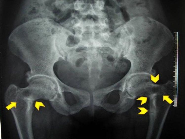
Tổn thương xương dẫn tới gãy giả xương đùi hai bên trên hình ảnh chụp X quang
2.3 Abnormalities in shape Fracture: The image shows a continuous loss of the bone margin, the two ends of the bone are displaced, the body is twisted, and the adjacent soft tissue is edematous. Post-healing changes caused by fractures cause the bone to enlarge or shrink. Lesions on film resemble rotting trees in osteomyelitis. In general, bone lesions on radiographs are very diverse, from the images of lesions on the film that the doctor can diagnose and guide the diagnosis of the disease. Bone X-ray is simple but has high value in the diagnosis of bone and joint diseases.
Currently, X-ray technique is a technique performed routinely at Vinmec International General Hospital to diagnose diseases. Depending on the pathology detected on the X-ray film, the doctor will give advice for each patient case. In order to improve the quality of medical examination and treatment, in addition to being fully equipped with modern medical systems and machines, and international standard X-ray machines, Vinmec also designs general health check-up packages suitable for patients. Each customer's age, gender and individual needs with a reasonable price policy, including:
Vip general health checkup package Special general health checkup package Standard general health checkup package With the above medical examination package, patients will have an in-depth examination with a team of specialists and perform diagnostic imaging methods such as X-rays, blood tests, and urine tests with the computer system. Modern hooks provide fast and accurate results.
The patient's examination results will be returned to the home. After receiving the results of the general health examination, if you detect diseases that require intensive examination and treatment, you can use services from other specialties at the Hospital with quality treatment and services. outstanding customer service.
Please dial HOTLINE for more information or register for an appointment HERE. Download MyVinmec app to make appointments faster and to manage your bookings easily.


