This is an automatically translated article.
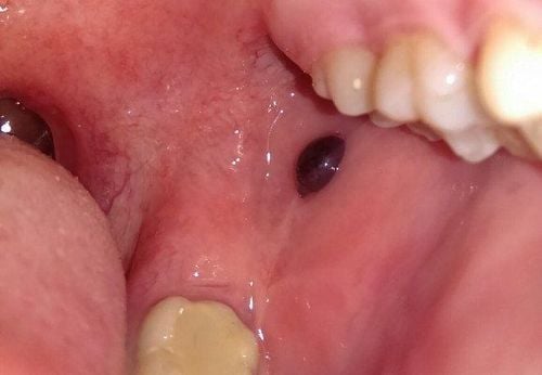
Hemangioma is a fairly common tumor, it can appear in many places on the body, usually under the skin, but it can also appear in the oral cavity. Are hemangiomas in the oral cavity dangerous and how are they treated?
1. Blood tumors in the oral cavity
Hemangioma is a tumor or abnormality under the skin or mucous membranes, usually congenital, it is composed of proliferation and dilation of blood vessels, usually capillaries, which are linked by affiliated organizations. There are also cases where hemangiomas are composed of a true cavernous tissue, like that of the erectile organ.
Histologically, there are two main types of hemangiomas:
Capillary hemangioma: This is the most common type of hemangioma, accounting for about 60%, with enlarged and dilated capillaries, but it has no proliferation of endothelial cells. Capillary hemangiomas consist of capillaries at different stages of development, some of which are hollow, some that are full, wide, and irregular in size compared to normal capillaries. Cavernous hemangioma: This type of hemangioma accounts for about 30% of the time, it has a structure similar to the erection organs consisting of small, blood-filled cavities that communicate with each other, and they often have a fibrous capsule that can press on Hard hold at the bottom. Sometimes these small cavities are separated from each other by collagenous walls that have lots of saggy tissue and lack of elastic. In cavernous hemangiomas, the capillaries are very dilated. Between these two types of hemangioma, there can be a combination on a single lesion. In addition, each of these hemangiomas can combine with other lesions, especially with lymphangiomas, to form hemangiomas or with other tissues such as muscle, bone, cartilage, etc.
Hemangiomas differ from vascular malformations in the following features:
Clinically: About 50% of hemangiomas appear at birth, the rest appear in the first month of life. Hemangiomas progress in 2 distinct stages: Right from the onset is a stage of rapid growth (tumor appears clearly, spreads and emerges on the surface of the skin or mucous membranes), lasts up to 6 – 2 years. 8 months after that will slow down and regress.

Trong miệng xuất hiện hạt đỏ có phải u máu trong khoang miệng không?
Histopathology: Hemangiomas have a strong hyperplasia of endothelial cells and an increase in the number of Mastocytes (playing a role in neovascular proliferation) in the proliferative phase, in the regressive phase of the number of cells. Mastocyte cells are normal but grow and infiltrate many fibrous and fatty tissues. Whereas in vascular malformations, the endothelial cells do not proliferate, they lie in a flat inner layer, which can change in the outer layers of the vessel.
On hematology: Hemangiomas can increase platelets, causing blood clotting in the tumor's lumen, while in vascular malformations, there are no blood disorders. Regarding hemodynamics, there is rapid blood flow in hemangiomas, while in vascular malformations, only the arteriovenous malformation has fast blood flow. Oral hemangiomas account for about 10%, it resembles intradermal hemangiomas, and can be flat, humpy, or tuberous hemangiomas. No tangled aneurysms were found in the oral mucosa.
Hemangiomas in the oral cavity often appear on the tongue, lips, cheeks, floor of the mouth, cleft palate, especially the soft palate, spreading to the tonsils combined with the uvula to form a hump, tubercular, very dangerous .
The mucous membrane covered with hemangiomas in the mouth can be from crimson to dark purple, it can be slightly bumpy or more, it easily causes bleeding and entanglement, affecting eating, drinking and speaking.
Oral hemangioma can also spread beyond the skin, after it invades submucosa, muscle, and subcutaneous fat. It usually grows in width, scattered in many places in the oral cavity.
Although a hemangioma in the mouth usually does not affect the aesthetics, it causes a lot of dysfunction because the tumor itself is entangled and causes bleeding and infection.
2. How to treat hemangiomas in the oral cavity?
Hemangiomas can be self-limiting, or stable, do not develop further, or continue to grow rapidly or slowly, may be life-threatening, or affect function, aesthetics, or not.
When a hemangioma has existed from birth until the child is 2-3 years old, it will usually continue to grow rapidly or slowly, but at least grow rapidly according to the person.
Depending on each case, the doctor will prescribe intervention or continue monitoring. When there are indications for interventional treatment of hemangiomas, there will be the following treatment methods:

Điều trị u máu trong khoang miệng bằng phương pháp đồng vị phóng xạ
Physics: Radioactive rays, radioactive isotopes, radium. Chemistry: Using sclerotherapy injections. Surgery ranges from minor surgeries such as grinding, scraping, tattooing, and staining. Surgery ranges from sclerotherapy, partial resection, to major surgery, total excision and reconstruction. Depending on each specific case, such as each disease, each location of the blood tumor, the doctor will advise you on the appropriate treatment method.
With hemangiomas in the mouth, the treatment direction is as follows:
Flat, localized hemangiomas, usually using surgical resection is the best. Although the aesthetic requirements are not as high as that of flat skin hemangiomas, this case needs to be functional because if the sutures are wrinkled, it will easily cause scarring, affecting eating and drinking, sometimes pulling the lips. nose. Hemangiomas in the mouth are tuberous or cause bleeding, so they need to be removed or used sclerotherapy.
Please dial HOTLINE for more information or register for an appointment HERE. Download MyVinmec app to make appointments faster and to manage your bookings easily.


