This is an automatically translated article.
The article is professionally consulted by Master, Doctor Le Xuan Thiep - Department of Diagnostic Imaging - Vinmec Ha Long International Hospital
There are many diagnostic imaging methods to help diagnose cardiovascular diseases such as ultrasound, angiography, CT Scanner, MRI, scintigraphy... In which, cardiovascular radiography is a method of applying X-rays to Image survey is used quite commonly today.
1. Heart Anatomy on Cardiovascular X-ray Film
The application of cardiovascular radiology is increasingly widespread in the diagnosis of heart disease. Normally, when taking a cardiac X-ray, there will be 3 basic positions: straight-back-anterior, left-tilt and oblique.
With each different imaging position, the anatomical landmarks of the heart are also conventions differently:
Post-anterior rectilinear position: The right border of the heart includes 3 arcs: superior vena cava, right atrium and inferior vena cava (often not obvious); The left side of the heart consists of 4 different arcs from top to bottom: the aortic arch, the pulmonary artery, the left atrium and the left ventricle. The left atrium alone may not be seen in people without cardiovascular disease. The left side position: The anterior border has 3 arcs including the right ventricle, the pulmonary trunk and the ascending aorta; The posterior border has two arches, the left ventricle and the left atrium. Oblique position: Includes front right and left front. Currently, this position of cardiovascular radiography is often indicated in cases where coronary imaging is required.
2. Cardiac X-ray image

Bóng tim trên x quang
2.1. Heart shadow The size of the heart shadow is the first feature to note when applying cardiac radiography to the diagnosis of heart disease. The large or small heart shadow is reflected through the heart-thorax index.
This index is the ratio of the maximum horizontal dimension of the heart shadow to the maximum width of the thorax (measured at the top of the diaphragm), which is normally about 0.5-0.55. When the cardiac-thoracic index on the radiograph changes, it suggests some of the following diagnoses:
Cardiothoracic index > 0.55 suggests an enlarged heart, seen in diseases such as heart failure, cardiomyopathy, abnormality. heart valve, pericardial effusion or mucinous tumor... Cardiothoracic index < 0.5 suggests a small heart, which can be seen in cases of cardiac anomalies, pericarditis, ventricular constriction or emphysema... 2.2 . Heart shape To diagnose heart disease, the heart shape on radiographs is considered an important feature:
Heart shaped like a water bottle (droplet) seen in pericardial effusion; Pathological ways of increasing the size of the right ventricle such as heart failure, the apex of the heart will rise higher than normal; Conversely, if the patient's left ventricle is large, the apex of the heart is lowered; Cardiomyopathies that cause thickening of the myocardium cause changes in the cardiac margins on cardiac radiographs, which are called defective margins; An enlarged aortic arch is seen in aneurysms or long-term hypertension; An enlarged pulmonary artery is seen in chronic obstructive pulmonary disease or an abnormal shunt from the left heart to the right heart. Normal pulmonary artery diameter is less than 1.5cm;
2.3. Changes in the size of the heart chambers Large left atrium: This sign suggests some of the following diagnoses of heart disease:
Wide main bronchus angle over 60 degrees; The left main bronchus is elevated; Enlarged left atrium on cardiac radiograph may be due to enlarged left atrium; Left atrial enlargement with a double border created by the left atrium with the right border of the heart. Right atrium enlargement: Suggested when the right atrium greater than 5.5 cm from midline increases convexity of the right lower border of the heart;
Left ventricular enlargement: Criteria for diagnosis of left ventricular enlargement on cardiac radiographs include:
The left ventricular margin is rounded at first, corresponding to the ventricular hypertrophy phase, when moving to the dilated phase, the left ventricle is long , straight out; The apex of the heart is downward; Hoffman Rigler sign (+). Right ventricular enlargement: Diagnostic criteria include apex elevation on straight film and retrosternal stenosis or cardiac shadow greater than one third of sternum height on lateral cardiac radiographs.
2.4. Pulmonary circulation

X quang tim mạch chuẩn đoán tăng áp động mạch phổi khi đường kính động mạch phổi gốc lớn hơn 15mm
Normally, pulmonary circulation on cardiac radiograph is distributed according to the rule of 1⁄3 when the number of blood vessels gradually decreases from 1⁄3 below to 1⁄3 above. Besides, the vascular diameter of the apex is smaller than that of the base of the lung, the ratio is about 0.5/1.0
Pulmonary hypertension (Plethora):
Usually, increased pulmonary circulation in common heart diseases is congenital heart disease. birth or shunt abnormality left to right; Criterion for the diagnosis of pulmonary hypervolemia on cardiac radiographs is that the large pulmonary artery in the umbilical region extends to the periphery with an increase in the diameter of the vessels at the apex and base of the distribution. At this time, the ratio of the apical and basal vascular apertures is 1.0/1.0. Oligaemia:
This abnormality is common in congenital heart disease tetralogy of Fallot (TOF). On X-ray, there will be 2 abnormally brightened lung fields. Pulmonary vessels have a small aperture, which is sometimes not seen on film. Pulmonary hypertension: Diagnosed when the diameter of the primary pulmonary artery is greater than 15mm.
2.5. Calcification Calcification on radiographs is a hyperattenuated spot (white), commonly seen in locations related to the heart such as:
Calcification of the coronary system; Closure of heart valves such as mitral valve to create an arc shape; Closure of the pericardium ; Closure of the aorta..
3. Issues to note when analyzing cardiac radiographs

Phân tích các tuần hoàn phổi là một trong các vấn đề cần lưu ý khi phân tích phim X quang tim-mạch
Evaluation of heart position, size of the heart shadow (cardio-thoracic ratio) Analysis of the dimensions of the chambers of the heart Location, size of the great vessels Analysis of the pulmonary circulation
4. Application of Cardiovascular X-ray
Application of cardiovascular radiographs to support the diagnosis of the following cardiovascular diseases:
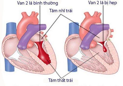
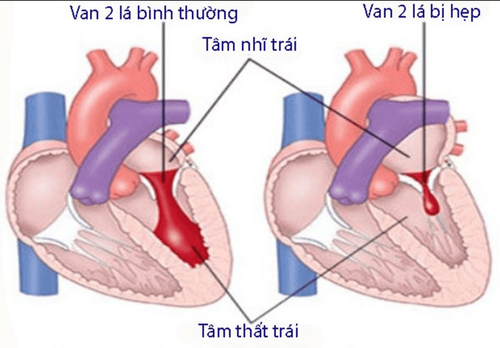
Mitral valve stenosis: Cardiovascular radiographs are characterized by pulmonary venous hypertension, left atrial enlargement and left atrial appendage while the left ventricle remains normal; Mitral regurgitation: There are images such as enlarged heart, enlarged atria and left ventricle. In addition, some other features may be encountered such as pulmonary venous hypertension (mild than mitral stenosis), mitral annulus calcification. Aortic stenosis : Usually difficult to diagnose on plain film; Aortic regurgitation: Enlarged heart ball, enlarged left ventricle and aortic arch; Ventricular septal defect: Cardiac radiographs will depend on the size of the orifice. If the stoma is small, most of the time there is no abnormality on the X-ray. When the stoma is larger, the heart shadow will become larger, accompanied by an increase in the size of the pulmonary artery. Atrial septal defect: Cardiovascular radiographs include thickening of the right atrium, enlargement of the right ventricle, and aneurysm of the pulmonary artery. Besides, the left ventricle is normal, the aortic arch is small.

Hẹp van hai lá
Tetralogy of Fallot: The apex of the heart is raised due to thickening of the right ventricle, a defect in the pulmonary artery, and the heart is shaped like a shoe. Left heart failure has images such as large, thickened and enlarged heart shadow, left atrium and left ventricle, pleural effusion, alveolar edema. Right heart failure is characterized by enlarged, enlarged right heart chambers. Pericardial effusion: Depends on the amount of fluid in the pericardium, if it is small, the precordial line is thicker than 2-3cm), the cardiac density and the density of the fluid are different. If the pericardial effusion is large, the heart shadow is enlarged symmetrically, the angle of the center of the diaphragm is small, the heart is watery, and the heart edges are not clear. Cardiovascular X-ray is a method of applying X-rays to survey images that is quite commonly used today. However, for accurate, clear and true image results, patients should go to reputable specialized medical centers for examination and implementation.
Vinmec International General Hospital has applied X-ray techniques in examining and diagnosing many diseases, including cardiovascular disease. X-ray technique at Vinmec is carried out methodically and according to standard procedures by a team of highly qualified medical professionals, modern machinery system, thus giving accurate results, making a significant contribution to diagnosis and staging of the disease.
If you have a need for medical examination by modern and highly effective methods at Vinmec, please register for an online examination.
Please dial HOTLINE for more information or register for an appointment HERE. Download MyVinmec app to make appointments faster and to manage your bookings easily.


