About

The Department of Diagnostic Imaging and Nuclear Medicine consists of two divisions: The Department of Diagnostic Imaging was formed after the establishment of Vinmec International General Hospital and is one of the spearheads in professional development orientation of the Hospital. The Nuclear Medicine Unit was formed at the end of 2017.

Why should you choose the Department of Diagnostic Imaging and Nuclear Medicine at Vinmec Times City International Hospital?

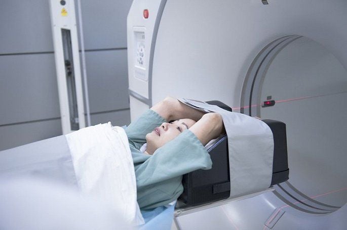
The Department of Diagnostic Imaging and Nuclear Medicine is a unit that is fully and synchronously equipped with the most modern and new generation machines in Vietnam. The department installed a 3D breast ultrasound machine to help add breast cancer screening facilities and the 3 Tesla GE Magnetic Resonator which helps to reduce noise. It is the second hospital in Hanoi equipped with new generation PET/CT and SPECT/CT machines, Hybrix angiography system, CT Angio system combines CT 640 slices with intelligent Angio system. We are proud that the Department of Diagnostic Imaging and Nuclear Medicine at Vinmec is the department equipped with the most modern and synchronous equipment in Vietnam and in Southeast Asia.

The department has a large number of staff members with 50 staff members, including 18 doctors (including 1 associate professor and 1 doctorate), who are regularly trained on-site or at other hospitals. short-term and long-term training courses at home and abroad. Training cooperation with Kantonsspital Winterthur Hospital (Switzerland) and Stanford University (USA), University of Sherbrooke (Canada) working together, exchanging expertise, regularly consulting with a team of experienced doctors of major faculties in Hanoi such as Bach Mai Hospital, Department of Diagnostic Imaging, Hanoi Medical University, Military Hospital 108.

The department has become a trusted address, in which doctors inside and outside the hospital put their confidence to refer patients to magnetic resonance imaging of brachial plexus lesions, brain tumors, pediatric cerebral palsy, CT scans of heart diseases Congenital complex in children, ultrasound measurement of elasticity of liver parenchyma, thyroid....

The good coordination between the Clinical departments, the Anesthesiology - Anesthesia Department and the Vinmec Department of Imaging - Nuclear Medicine has ensured the good and safe performance of all Magnetic Resonance and Radiography examinations. Computer class, PET/CT, SPECT/CT Angio - Hybrix solve problems from diagnosis to treatment intervention for complex diseases of cancer, deformities in adults. As in very young children, Magnetic resonance imaging for children from 3 days old with malformations of the spinal canal, skull, biliary tract, ...

Perform routine biopsy techniques, learn to aspirate cells. This is considered the gold standard in the diagnosis of some diseases, especially the assessment of cirrhosis in congenital biliary atresia, cancer diseases.

The department is promoting the field of intervention with the means of two-sided DSA machine, Hybrit angiography system, 640-slice CT-Angio system, which can receive and treat diseases such as primary hepatocellular tumor, tumor uterine fibroids, prostate fibroids, retinoblastoma... by techniques such as embolization, implementation of brain aneurysm node intervention, spinal vascular malformations, bronchial vasodilation, renal vascular malformations. In addition, performing radiofrequency ablation (RFA) for liver tumors, benign thyroid tumors, etc.

Professional achievements:

  • Routine diagnosis of liver fibrosis by elastography of liver tissue.
  • Application of multi-sequence CT - Scan system to accurately assess before organ transplantation: normal or anatomical variation of organs and blood vessels in liver and kidney transplantation.
  • Digestive fluid erasure application along with biliary pulse sequence to evaluate biliary anatomical changes. Assess the degree of steatosis, iron contamination in the liver.
  • Evaluation of brachial plexus injury in infants and adults.
  • Evaluation of axonal damage in cerebral palsy, brain tumor.
  • Evaluation of magnetic resonance lesions in cerebral palsy, autism.
  • Evaluation of metabolic damage in autism in children.
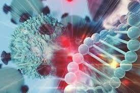
  • Application of PET/CT in oncology.
  • Collaborate with experts in the treatment of brain aneurysms, arterial thrombosis.

Working position: The Department of Diagnostic Imaging is located on the 1st floor of the Hospital

Our Doctors
Facilities

The Department of Diagnostic Imaging and Nuclear Medicine is synchronously equipped with the most modern and advanced equipment compared to domestic and regional hospitals:

  • 9 color Doppler ultrasound rooms.
  • 1 breast ultrasound room.
  • 3 digitized X-ray rooms – DR (1 full-axis, 1 intensifier and 1 synthesizer)
  • 2 portable DR X-ray machines
  • 2 multi-slice computed tomography rooms with receiver (1 384-slice machine (replace 128-slice) and 1 256-slice dual-energy machine)
  • 2 Magnetic resonance imaging rooms (1 machine 3 Tesla Siemens and 1 machine 3 Tesla GE)
  • 1 interventional angiography room with 2 planes, 1 room for Hybrid vascular intervention, 1 room for CT-Angio intervention.
  • 1 bone mineralometer.
  • 1 PET/CT 128 series GE machine.
  • 1 SPECT/CT GE machine.
Services
  • The Department of Diagnostic Imaging and Nuclear Medicine provides all diagnostic and treatment services for domestic and foreign customers.
  • Periodic health examination for companies and organizations.
  • Meeting requirements of high-tech diagnostic imaging services from other medical centers.
  • Screening for cancer, assessing the stage of cancer, etc.
  • Services:
  • Ultrasound.
    • General abdominal ultrasound, ultrasound of mammary gland, testicles, thyroid gland, etc.
    • Ultrasound for diagnosis of gastroesophageal reflux disease.
    • Soft tissue and musculoskeletal ultrasound.
    • Ultrasound of blood vessels.
    • Elastography of liver, thyroid gland, mammary gland.
    • Ultrasound for guiding biopsy, abdominal paracentesis, pleural paracentesis, etc.
    • Obstetrics and gynecology ultrasound, etc.
  • Radiography (X-ray).
    • Conventional X-ray of chest, bone, joint, cranium, sinus, unprepared abdomen, unprepared urinary tract, etc.
    • Full-length lower limb X-ray.
    • X-ray of stomach-esophagus, small intestinal circulation, colon, urinary tract with contrast, uretero pyelographie retrograde, fistula, etc.
    • Hysterosalpingography (no pain)
    • Panoramic dental X-ray
    • Mammography.
    • Bone densitometry
  • Computerized tomography (CT)
    • Routine CT of brain, sinus, spine, etc.
    • CT of cerebral stroke (multiphase, perfusion).
    • Chest CT with contrast, high resolution.
    • CT of abdomen–pelvis, urinary tract.
    • Full body CT for assessing cancer, screening metastasis.
    • Coronary angiography.
    • Angiography of abdominal and thoracic aorta, extremity angiography.
    • 3D bone-shape reconstruction.
    • Spectral CT: assessing metastatic lesions in cancer, assessing stone composition (uric acid stones or not), assessing monosodium urate crystals in  joints of patients with Gout, low-volume contrast CT angiography, etc.
    • CT for guiding biopsy.
  • Magnetic Resonance Imaging (MRI)
    • In accordance with the standard protocols.
    • MRI of brain, pituitary gland, orbit, intracranial blood vessels, carotid artery, etc.
    • MRI of stroke.
    • Tractography, function MRI.
    • MRI of cervical thoracic and lumbar spine
    • MRI of joints: shoulder joint with intra-articular injection, knee joint, wrist joint, phalangeal joint, elbow joint, hip joint, etc.
    • MRI of soft tissue of the neck and other parts.
    • MRI of brachial plexus
    • MRI of abdomen, biliary tract, pelvis (uterus – adnexa, prostate, etc.), anal fistula.
    • Dynamic pelvic floor MRI (Defeco).
    • MRI of fetus, placenta.
    • Breast MRI.
    • Heart MRI.
    • Blood vessel MRI
    • MRI under general anesthesia
  • Intervention unit.
    • Ultrasound for guiding biopsy of breast, thyroid gland, liver, soft tissue, etc.
    • CT for guiding biopsy of lungs, liver tumors, etc.
    • RFA of benign liver tumors, thyroid tumors.
    • Angiography with 2-plane DSA machine for diagnosis of vasculopathy.
    • Embolization for cerebral aneurysm, cerebrovascular malformation, transarterial chemoembolization (TACE) for liver tumors, embolization for hemoptysis, gastrointestinal bleeding, renal artery stenosis or renal arteriovenous malformation, uterine fibroids, etc.
    • Selective internal radiation therapy (SIRT) for liver cancer.
    • Diagnostic techniques and treatment for mammary gland diseases: Treatment for benign mammary tumors with negative pressure therapy device (lumpectomy with vacuum-assisted device Mammotome). Mammotome device-guided breast biopsy. Ultrasound and Mammotome device-guided hookwire placement.
    • Coordination in treatment for tendon and joint diseases by ultrasound-guided corticoid or PRP injections.
  • Nuclear Medicine Unit:
    • PET/CT for diagnosis of tumors.
    • PET/CT for evaluation of autism-cerebral palsy.
    • PET/CT for diagnosis of aphrenia.
    • PET/CT for diagnosis of Alzheimer's disease.
    • PET/CT for diagnosis of epilepsy.
    • PET/CT for diagnosis of dementia.
    • PET/CT for diagnosis of cardiovascular diseases.
    • PET/CT for diagnosis of myocardial vitality
    • PET/CT for diagnosis of infection.
    • PET/CT for radiation therapy simulation.
    • PET/CT for simulation of intensity modulate radiation therapy. 
    • Myocardial perfusion imaging with SPECT/CT, tagged red blood cell imaging for diagnosis of hepatic hemangioma.
    • Thyroid scintigraphy.
    • Kidney scintigraphy.
    • Bone scintigraphy, etc.
    • RFA of benign liver tumors, thyroid tumors.
    • Selective internal radiation therapy (SIRT) of liver cancer.
    • Breast biopsy with negative pressure therapy device, hookwire placement, marker placement, etc.
Languages spoken (2)

You have been taken care of at Diagnostic imaging and nuclear medicine department? You can share your experience and feedback here.

Vinmec guarantees absolute confidentiality with your email address and personal information.

  • Attitude:
  • Wait time:
  • Hygiene:
Yes No

Thank you for your feedback!

Your feedback will be used to help Diagnostic imaging and nuclear medicine department improve quality and service.

Please continue to share your experience and feedback in the future!

Chọn bác sĩ và giờ bạn muốn đặt khám từ lịch dưới đây. Để được tư vấn chọn bác sĩ, bạn có thể chat với chúng tôi trên trang web này hoặc gọi điện cho chúng tôi theo số trong Hotlines.

Map detail