This is an automatically translated article.
Posted by Master, Doctor Nguyen Quang Duc - Department of Diagnostic Imaging and Nuclear Medicine - Vinmec Times City International General HospitalLiver hemangioma with SPECT/CT machine is a very useful method with high accuracy to identify benign hemangioma lesions in the liver parenchyma. This technique is performed routinely at Vinmec Times City International General Hospital.
1. What is liver hemangioma?
Hemangiomas are benign tumors of the liver. In most cases, liver hemangiomas cause no symptoms and do not require any treatment. However, there are some malignancies in the liver that need to be treated with similar manifestations of liver hemangiomas, so a differential diagnosis with liver hemangiomas is necessary.2. Symptoms of hemangiomas in the liver
Most liver hemangiomas cause no symptoms, so it is only discovered by chance during physical examination, liver ultrasound or computed tomography, magnetic resonance imaging, etc.
Hemangiomas may be present. one or more blocks. Some large hemangiomas can cause symptoms but are very vague such as:
Feeling uncomfortable, full stomach Nausea Loss of appetite... Some liver hemangiomas are large, located close to the liver. The chest wall can be prone to rupture when trauma causes intra-abdominal bleeding.

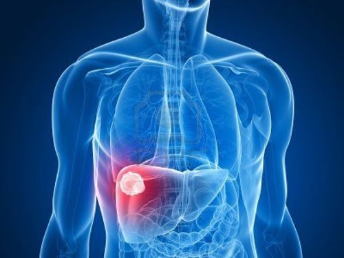
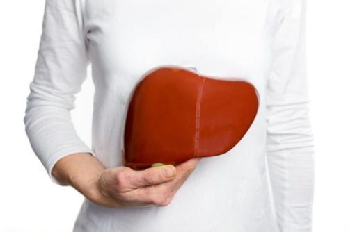
Hình ảnh minh họa khối u máu trong gan được chẩn đoán bằng CT
3. How are liver hemangiomas diagnosed?
Liver hemangiomas are often detected by imaging techniques such as ultrasound, computed tomography (CT), magnetic resonance imaging (MRI). However, SPECT/CT scan is a diagnostic method with very high accuracy up to nearly 100%, especially for liver hemangiomas over 1 cm in size. As a result, patients avoid unnecessary diagnostic investigations such as biopsies, thereby limiting costs, saving time and minimizing pain and complications for the patient.

Hình ảnh u máu gan trên CT (bên trái) và SPECT/CT (phải)
4. What to prepare when going to the liver hemangioma scan at Vinmec Times City hospital?
Bring all old records (results of medical examination, ultrasound, computed tomography...). Notify the status of pregnancy, lactation (if any) to medical staff before performing the service.
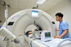
5. Why should liver hemangioma scan at Vinmec Times City Hospital?
Vinmec uses the SPECT/CT Discovery NM/CT 670 Pro system with the most modern 16-sequence CT of the world's leading medical equipment company GE Heathcare (USA), for high quality images, helping to diagnose diseases early reasons to be investigated. Because the image is combined between SPECT and CT images, it is able to accurately locate the lesion as well as provide more useful information from CT to help increase the diagnostic value of the disease. Vinmec hospital's high quality service will satisfy customers. Vinmec's medical staff are experienced, well-trained at home and abroad, so they can advise and provide maximum support to customers during the shooting process, even for foreign customers. For advice on liver hemangiomas, please contact the nuclear medicine unit hotline, Vinmec Times City International General Hospital: 0979 866 368 or register online HERE