This is an automatically translated article.
The article was professionally consulted by Specialist Doctor I Do Van Manh - Emergency Resuscitation Doctor - Emergency Resuscitation Department - Vinmec Ha Long International HospitalIn the treatment of patients with advanced acute respiratory distress syndrome, prone position artificial ventilation has many effects such as improving blood oxygen, reducing the risk of lung injury due to mechanical ventilation,...
1. Significance of artificial ventilation in prone position for patients with acute lung injury?
1.1 Severity of acute respiratory distress syndrome Acute respiratory distress syndrome (ARDS) is a severe, rapidly progressive medical condition. This is a critical condition in terms of gas exchange function and causes hypoxia in the body's arterial blood, with an overall mortality rate of 30 - 60%, of which the death rate of severe patients is up to 80%. . The characteristic manifestation of the disease is diffuse damage to the alveolar capillary membrane, causing prolonged refractory hypoxemia.
Therefore, it is necessary to perform first aid for acute respiratory failure in accordance with the principles and conduct it urgently to promptly save the patient's life and not cause complications due to emergency resuscitation.
1.2 Effect of prosthetic ventilation in prone position Pronation method of artificial ventilation in the treatment of patients with acute respiratory failure is significant:
Effects in lung mechanics:
Increases lung compliance; Synchronized ventilation between the dorsal and sternal lung areas; Open collapsed alveoli, increase alveolar ventilation. Improve blood oxygen and reduce shunt in the lung:
prone position artificial ventilation to reduce shunt, adjust ventilation/perfusion ratio. Helps protect the lungs:
The prone position helps to more evenly distribute lung density, intrapulmonary shunts, transpulmonary pressure and pulmonary ventilation. Therefore, this position helps the lungs to be less stretched during mechanical ventilation and distributes pressure more synchronously in the lung areas. The prone position of artificial ventilation also reduces excessive alveolar dilation and cyclic collapse of the collapsed alveoli, and also reduces the production of inflammatory cytokines.
prone position artificial ventilation to reduce mechanical ventilation time, shorten hospital stay and reduce mortality. However, prosthetic ventilation should be introduced in the prone position as early as 24 hours, in critically ill patients with PaO2/FiO2 ≤ 150 mmHg.
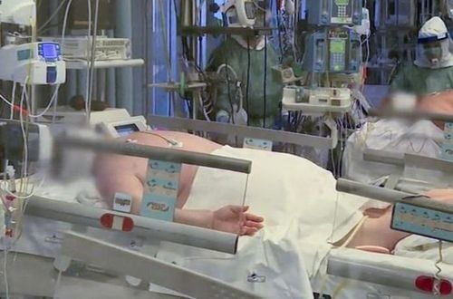
2. Procedure for artificial ventilation in prone position
2.1 Some important notes Time to conduct prosthetic ventilation in prone position: After artificial ventilation according to ARDS Network 12 - 24 hours without improvement in blood gas, PaO2/FiO2 ratio ≤ 100 mmHg or PaO2 /FiO2 ≤ 150 mmHg but tends to continue to decrease, PEEP ≥ 5 cmH2O, FiO2 ≥ 60%; Time of artificial ventilation in prone position: 16 - 17 hours/day when blood oxygen improves (PaO2/FiO2 increases by more than 20 mmHg compared to the time before prone position) and no dangerous complications occur. If after 6 hours of supine supine, the PaO2/FiO2 ratio < 150 mmHg, the patient should continue artificial ventilation in the prone position for the 2nd or 3rd time,...; When to stop the artificial ventilation in the prone position: When the patient does not respond to the prone position such as: Blood oxygen does not improve after 6 hours of lying on the stomach, then switch to the supine position, continue mechanical ventilation as when prone; When the occurrence of dangerous events such as obstruction, extubation of the endotracheal tube, hypotension (mean blood pressure <60 mmHg after increasing the dose of vasopressors without response), circulatory arrest. The patient was transferred to the supine position, the doctor handled the complications such as emergency circulatory arrest, re-intubation, pleural opening for air drainage,...; The patient is no longer indicated for artificial ventilation in the prone position: After the patient changed from prone to supine position for 6 hours, PaO2/FiO2 ≥ 150 mmHg with PEEP ≤ 10 cmH2O, FiO2 ≤ 60%. The patient continued to receive artificial ventilation in the supine position. 2.2 Perform artificial ventilation in the prone position Preparing to change the patient's position:
Medical staff: Including 3 people who are doctors or nurses. 1 person to stand on the patient's head to raise the patient's head, keep the ventilator wire and the IV catheter from slipping; 2 people stand on either side of the bed to rotate the patient; Patient: Suctioned sputum, lying on the head position; continue mechanical ventilation during position change; maintain the use of sedatives, muscle relaxants; remove the urine bag; Technical facilities: 1 bed sheet, 4 soft pillows, 3 monitor electrodes, firmly fixed the ends of the ventilator connecting tube, endotracheal tube, nasogastric tube, IV catheter and other lines other transmission. Change the patient's position from supine to prone:
Pull the patient to one side of the bed (on the same side as the IV catheter to prevent the catheter from getting entangled in the patient's neck when changing positions); roll old bed sheets tucked under the patient's back; Spread half a new bed sheet on top of the bed. Keep the patient's arm straight, pressed close to the body; Turn the patient to a side lying position, remove the old bed sheet, move the electrocardiogram monitoring electrode to the patient's back corresponding to the positions in front of the chest; Turn the patient into a prone position, tilt the head to one side, and pull and spread the new bed sheet. Next, place a soft pillow under the patient's face, hips, chest and legs, and adjust the head of the bed to a height of 30°. At the same time, pay attention to change the patient's head position to the sides every 2-3 hours. When changing, 1 person lifts - rotates the patient's head to the opposite side, 1 person supports the ventilator, endotracheal tube and nasogastric tube; When transferring the patient from the prone position to the supine position, do the same: Move the patient to the edge of the bed on the same side as the catheter, place the patient in a supine position, remove the electrodes on the back to move to the front of the chest, and then place the patient in a supine position. The patient lies on his back and installs a monitor. Artificial ventilation while the patient is prone:
Maintain the ventilator with breathing mode, the same parameters as when the patient is lying on his back; The patient was opened with CPAP 40 cmH2O for 40 seconds - similar to when lying on the back; Adjust ventilator in prone position according to goal: Blood oxygen maintain PaO2 at 55 - 80 mmHg or SpO2 at 88 - 95%, maintain Pplateau lb 30 cmH2O, maintain pH from 7, 25 - 7.45, I:E ratio from 1:1 to 1:3.
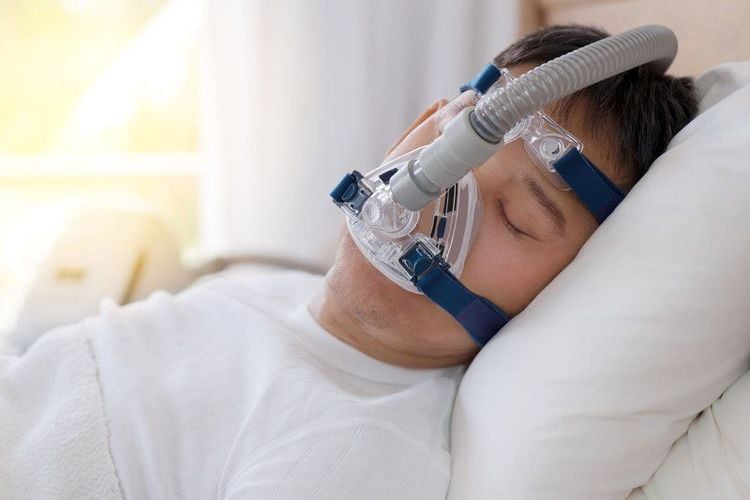
Artificial ventilation in the prone position in acute lung injury should be performed according to standard procedures to improve treatment efficiency and reduce the risk of complications for the patient. In addition, to help increase the survival chances of patients with acute lung injury, it is necessary to choose an emergency medical facility with a system of modern and reputable equipment.
Currently, Vinmec International General Hospital is one of the leading hospitals in the country, trusted by a large number of patients for medical examination and treatment. Not only the physical system, modern equipment: 6 ultrasound rooms, 4 DR X-ray rooms (1 full-axis machine, 1 light machine, 1 general machine and 1 mammography machine) , 2 DR portable X-ray machines, 2 multi-row CT scanner rooms (1 128 rows and 1 16 arrays), 2 Magnetic resonance imaging rooms (1 3 Tesla and 1 1.5 Tesla), 1 room for 2 levels of interventional angiography and 1 room to measure bone mineral density.... Vinmec is also the place to gather a team of experienced doctors and nurses who will greatly assist in diagnosis and detection. early signs of abnormality in the patient's body. In particular, with the space designed according to 5-star hotel standards, Vinmec ensures to bring the patient the most comfort, friendliness and peace of mind.
Please dial HOTLINE for more information or register for an appointment HERE. Download MyVinmec app to make appointments faster and to manage your bookings easily.


