This is an automatically translated article.
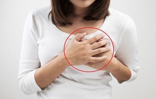
The article is professionally consulted by Master, Doctor Ton Nu Tra My - Department of Diagnostic Imaging - Vinmec Central Park International General Hospital.A breast lump is a mass or area of swollen breast tissue that may or may not be palpable. When a breast lump is detected, you should see a specialist as soon as possible.
1. How is a breast tumor diagnosed?
Most breast lumps are benign (not cancerous). To evaluate breast tumors, doctors often have to specify different imaging techniques. You may need one or more of the following imaging techniques:Mammography, mammogram, mammogram. A mammogram is an imaging method that uses low-dose X-rays to see inside the breast for the early diagnosis of breast cancer. It is the oldest and first used imaging tool in breast cancer screening.
Today, tomosynthesis is also known as three-dimensional (3-D) mammography. Tomosynthesis creates multiple images of the breast from different angles and reconstructs or "composites" into a set of three-dimensional images. Early studies show that this method is able to detect more breast lumps, especially breast lumps in women with dense breast tissue.
Breast ultrasound: is a technique that uses sound waves to create images of structures inside the breast. During ultrasound, the doctor uses a transducer (transducer) close to the skin, the transducer has the function of both transmitting and receiving ultrasound waves. During an ultrasound, the crystals inside the transducer emit ultrasound waves that travel inside the body. Tissues, bones and body fluids - partly absorbing or transmitting - partly reflect the sound waves and return to the transducer. The transducer receives the feedback sound waves, sends this information to the processor, after analyzing the feedback signals by image processing software and algorithms, combines the information to build and reconstruct into ultrasound images that help doctors see structures inside the breast, thereby detecting breast lumps.
Breast Magnetic Resonance: is an imaging technique used to detect breast cancer and other abnormalities in the breast. Magnetic resonance imaging of the breast is usually performed when the doctor needs more information about the extent of the disease that other imaging methods such as mammograms (mammograms) and ultrasound of the breast cannot respond to or are used. used to screen for breast cancer in women at high risk of breast cancer (For more information, please see “breast cancer risk”)
After performing the techniques If the radiologist concludes that the tumor is benign, then you will not need to do any more tests. However, doctors recommend that you continue to have regular breast exams to see if the breast lump has changed, stabilized, or disappeared.
If the imaging techniques above do not clearly show whether the tumor is benign or malignant, you may need a biopsy to determine the nature of the breast lump
There are many types of biopsies, specifically Fine-needle aspiration cytology (FNA): is a technique that uses a fine needle under ultrasound guidance to penetrate the breast tumor, apply negative pressure, and withdraw the contents contained within the tumor. for cytological examination. Core biopsy (CNB) under ultrasound guidance: A technique that uses a needle with a larger core than that of fine needle aspiration cytology, under ultrasound guidance to take tissue samples from the tumor for testing. X-ray-guided breast biopsy (Stereotaxic): is a technique of taking breast tissue samples under the 3-dimensional positioning of a mammogram machine for histopathological examination. MRI-guided biopsy (MRI-guided biopsy): is a technique to take breast tissue samples under the 3-dimensional positioning of the breast magnetic resonance machine for histopathological examination.
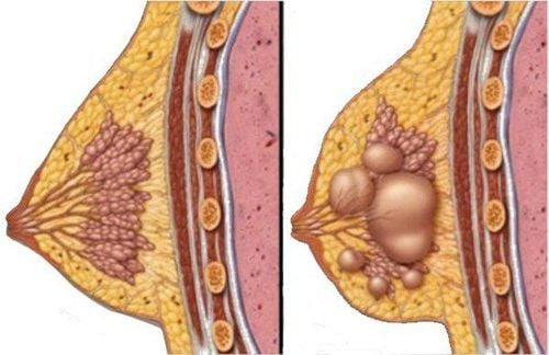
U nang vú có thể gây ra khối u ở vú
2. Results of breast tumor assessment
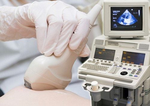
Siêu âm vú giúp chuẩn đoán và đánh giá khối u vú
If the diagnosis is suspected breast cancer after a physical examination and mammogram, but the biopsy shows benign tissue, you will be referred to a more specialized doctor for further advice.
If the breast lump is cancerous, you will be referred for an examination and consultation with an oncologist for staging and treatment planning. Treatment options depend on the stage of the cancer and the type of breast cancer.
At Vinmec International General Hospital, there is a Breast Cancer Screening Package, which helps customers screen and detect breast cancer early even when there are no symptoms. When registering for the Breast Cancer Screening Package, customers will receive:
Examination and consultation with an oncologist. Breast cancer screening by bilateral breast ultrasound and mammogram. Vinmec has become a prestigious address in breast cancer screening with:
A team of highly qualified and experienced doctors. Comprehensive professional cooperation with domestic and international hospitals: Singapore, Japan, USA, etc. Comprehensive treatment and care for patients, multi-specialty coordination towards individualizing each patient. Having a full range of specialized facilities to diagnose the disease and stage it before treatment: Endoscopy, CT scan, PET-CT scan, MRI, histopathological diagnosis, gene-cell testing, .. There are full range of mainstream cancer treatment methods: surgery, radiation therapy, chemotherapy, stem cell transplant...
Please dial HOTLINE for more information or register for an appointment HERE. Download MyVinmec app to make appointments faster and to manage your bookings easily.
Reference source: mayoclinic.org; radiologyinfo.org; healthline.com