This is an automatically translated article.
The article is professionally consulted by Master, Doctor Nguyen Thi Mai Anh - Radiologist - Department of Diagnostic Imaging and Nuclear Medicine - Vinmec Times City International Hospital. Doctor Nguyen Thi Mai Anh has nearly 10 years of experience in the field of diagnostic imaging, especially in imaging breast and thyroid cancer.Liver ultrasound is the most common imaging technique used to diagnose fatty liver today. Not only helping to detect disease, liver ultrasound also helps evaluate the degree of fatty liver, thereby helping doctors to have effective treatment for each patient.
1. What is fatty liver disease?
Fatty liver is caused by the accumulation of too much fat in liver cells. In normal people, the amount of fat in the liver is very low, only 2-4%. Mild fatty liver when the amount of fat in the liver accounts for 5-10%, moderate steatosis when the amount of fat in the liver accounts for 10-25%, severe steatosis when the amount of fat in the liver exceeds 30%.
Gan nhiễm mỡ xảy ra khi mức độ nhiễm mỡ vượt qua quy định cho phép
2. Causes of fatty liver
The causes of fatty liver can be divided into 2 main groups that are alcoholic and non-alcoholic fatty liver:2.1 Alcoholic fatty liver
Chronic alcoholism increases the synthesis and breakdown of fat in the liver, leading to fat accumulation in the liver.2.2 Non-alcoholic fatty liver
Non-alcoholic fatty liver includes many causes such as: Overweight, obesity, diabetes, hyperlipidemia, side effects of some drugs such as Aspirin, steroids,...Overweight, obesity: When the body regularly supplies fat beyond the threshold the body can absorb, there will be a phenomenon of accumulation, not fully metabolized, causing the risk of fatty liver. The more severe the obesity, the more severe the degree of fatty liver. Diabetes: Fatty liver is a very common condition in patients with type II diabetes due to a disorder of fat metabolism. Hyperlipidemia: Hyperlipidemia is often accompanied by a relatively high rate of fatty liver. Fatty liver rarely has obvious symptoms, the patient may feel a little warm and uncomfortable in the abdomen. When fatty liver condition progresses, the amount of fat in the liver is too much, the patient may have symptoms such as: Nausea, bloating, poor appetite, weight loss, fatigue, jaundice,... If detected and treated promptly, fatty liver will progress to steatohepatitis, cirrhosis and liver cancer, threatening the patient's life.

Người béo phì tăng nguy cơ bi gan nhiễm mỡ
3. Image of fatty liver on ultrasound
3.1 What is liver ultrasound?
To diagnose fatty liver disease, the doctor will directly examine the patient, ask about the medical history, assign the patient to perform blood tests and perform imaging techniques, etc. With low cost, safety, high sensitivity and specificity in detecting fatty liver, liver ultrasound is the most commonly used imaging technique in clinical practice.When performing an ultrasound of the liver, the doctor will use an ultrasound machine with an instrument that emits ultrasound waves to the right side of the patient's abdomen to observe the image of the liver.
These images are displayed on the screen, helping the doctor to detect liver damage. Liver ultrasound can delineate lobes, segments, and subsegments. Liver injury was detected on ultrasound based on the relationship of the aorta to the inferior, superior, and portal veins of the liver.
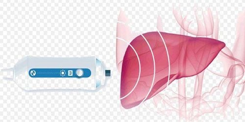
Siêu âm gan giúp đánh giá tình trạng gan nhiễm mỡ
3.2 Image of fatty liver on ultrasound
Fatty liver disease detected on ultrasound is mainly based on symptoms such as increased brightness in different areas of the liver or increased brightness of liver tissue causing the border of vascular structures to become blurred.On ultrasound images, liver morphology will appear scattered or concentrated light spots in areas. This situation will be different depending on the degree of liver fat in each person. When the liver is fatty, the sonographer will not see or clearly see the external vascular system on the liver.
In addition, normal liver echogenicity is equal to or slightly increased compared to renal cortex or spleen. When the liver is fatty, the echogenicity of the liver is superior to that of the renal cortex and spleen, and there is attenuation of ultrasound waves, loss of diaphragm clarity, and poor delineation of intrahepatic structures.
3.3 Assess the degree of fatty liver on ultrasound of fatty liver
The doctor can assess the degree of fatty liver based on the phenomenon of increasing the brightness of the liver parenchyma on the liver ultrasound image as follows:Level 1: the liver is mildly hyperechoic, the level of sound absorption has not changed. Significantly, the diaphragm and the margins of the intrahepatic veins are still identifiable. Grade 2: increased diffuse echogenicity and aspiration, the ability to identify the margins of veins in the liver and diaphragm has been greatly reduced. Grade 3: markedly increased echogenicity and aspiration, no longer recognizing the border of the veins in the liver, diaphragm, part of the liver parenchyma in the right posterior segment of the liver on the subcostal view. Fatty liver ultrasound helps doctors assess the extent of liver damage, thereby giving appropriate treatment for each patient.

Siêu âm gan là phương pháp chẩn đoán hình ảnh an toàn, không gây xâm lấn
4. Accuracy of fatty liver ultrasound
Due to many advantages such as high safety, low cost, high sensitivity and specificity, ultrasound of fatty liver is the most commonly used method today.However, because ultrasound is an imaging method that depends on the doctor's eyes, it is highly subjective. Diagnostic results may be inaccurate if the sonographer is inexperienced or the machine is set up incorrectly.
Besides, liver ultrasound does not distinguish between simple fatty liver or fatty degeneration causing hepatitis. For an accurate diagnosis, the doctor will assign the patient a CT or MRI scan,..
Please dial HOTLINE for more information or register for an appointment HERE. Download MyVinmec app to make appointments faster and to manage your bookings easily.