This is an automatically translated article.
The article is professionally consulted by Master, Doctor Nguyen Thuc Vy - Doctor of Diagnostic Imaging - Department of Diagnostic Imaging - Vinmec Nha Trang International General Hospital.Liver ultrasound is a non-invasive, safe and effective imaging technique in the diagnosis of certain liver diseases and liver damage. However, to ensure accurate liver ultrasound results, patients should note some points outlined below.
1. What is ultrasound?
Ultrasound is a safe, non-invasive, painless diagnostic technique that does not use radiation. This is a method with high diagnostic value, convenience, and low cost. So far, there have been basically no harmful effects of ultrasound on the human body. Therefore, it can be repeated many times for diagnosis, monitoring and combined treatment procedures.Ultrasound scanner includes computer control panel, video display and attached transducer. The transducer is a small handheld device resembling a microphone. The transducer emits high-frequency sound waves, or ultrasound, into the body and then picks up the echoes back.
The sonographer applies a small amount of gel to the area to be examined and places the transducer there. The gel allows sound waves to travel back and forth between the transducer and the area being examined. The ultrasound image is immediately visible on the display. The computer creates an image based on loudness (amplitude), intensity (frequency), and the time it takes for the ultrasound signal to return to the transducer. It also takes into account the type of body structure or tissue through which the sound travels. In medicine, ultrasound is used to detect changes in the appearance of organs, tissues, and vessels and to detect abnormal masses, such as tumors.

Siêu âm gan là kỹ thuật chẩn đoán hình ảnh không xâm lấn, an toàn, hiệu quả trong chẩn đoán một số bệnh lý gan, tổn thương gan nhất định.
2. Meaning of liver ultrasound
Liver ultrasonography can be performed separately or as part of a general abdominal ultrasound technique. This technique helps to delineate segments, lobes, and subsegments of the liver, based on the relationship to the aorta, inferior vein, portal vein, and hepatic vein, to detect pathologies and lesions. of the liver.The images displayed on the ultrasound help the doctor diagnose certain diseases or liver damage such as:
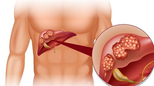
Fatty liver: Observation on ultrasound shows that part or all of the liver parenchyma is bright Cirrhosis: Patients with cirrhosis in the early stages have an increased liver size at first, then a smaller liver later. In the late stages of cirrhosis, ultrasound can also show the liver as a small nodule less than 1cm in diameter. In addition, ultrasound also helps detect consequences of cirrhosis such as ascites, varicose veins, and enlarged spleen. Liver cancer: Ultrasound is one of the important tests to help diagnose liver cancer Hepatitis: On ultrasound, liver lesions are observed, liver size increases, but the image of liver parenchyma has not changed. Obvious Liver abscess Liver fluke Other hepatobiliary diseases
3. Does liver ultrasound require fasting?
Usually, liver ultrasound does not require fasting. However, to better examine the bile ducts in and out of the liver, your doctor may ask you to fast for 8 to 12 hours before the ultrasound. Because if the ultrasound after eating, the gallbladder collapses, it will be difficult to assess the damage inside the lumen and wall of the gallbladder, in addition, when the stomach contains too much food in the head of the pancreas, the end of the common bile duct and part of the left liver. difficult to see on ultrasound.
Thông thường, bác sĩ thường sẽ yêu cầu bạn nhịn ăn trong vòng 8 đến 12 giờ trước khi siêu âm
4. Liver ultrasound procedure
Before your liver ultrasound, you may be asked to change into a hospital gown and remove any jewelry. You will be asked to lie on your back on an examination table. The sonographer applies a small amount of gel to your abdomen. The sonographer gently presses the transducer into your abdomen, moving it back and forth. The device sends signals to a computer, which creates images that help your doctor see the structure of your liver. An ultrasound exam usually takes about 10 to 30 minutes to complete. It is usually painless. However, you may have some temporary discomfort if the technician presses on a painful or tender area. You should be able to return to normal activities immediately after a liver ultrasound.5. Several factors can affect the results of liver ultrasound
Several factors or conditions can affect ultrasound results, including:Severe obesity Food left in the stomach Barium (a liquid you drink to do some tests that helps your doctor see your stomach and digestive tract) left over in your intestines from a previous test. Excess intestinal gas.
Please dial HOTLINE for more information or register for an appointment HERE. Download MyVinmec app to make appointments faster and to manage your bookings easily.