This is an automatically translated article.
The article is professionally consulted by MSc, BS. Dang Manh Cuong - Radiologist - Radiology Department - Vinmec Central Park International General Hospital. The doctor has over 18 years of experience in the field of ultrasound - diagnostic imaging.X-ray is one of the imaging methods that bring very good results in the diagnosis of joint diseases, including radiographic technique of the elbow joint.
1. What is an elbow joint?
The elbow is the joint between the arm and the forearm, an important bridge creating cohesion and rhythmic movement of the hand. When the elbow joint is painful, the hand will have a lot of difficulty in moving.2. What is an X-ray of the elbow joint?
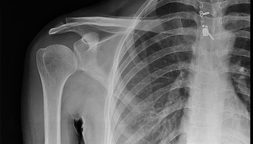
X-ray of the straight elbow joint is one of the simple techniques indicated when the patient is suspected of having bone and joint diseases.With the operating mechanism of X-rays, the movements and images inside the body will be shown quite clearly on the film, creating favorable conditions for the doctor to diagnose the disease without having to interfere with the procedures. complex.

3. X-ray of straight elbow joint in which case?
Radiographs of the elbow joint are recommended regularly to detect internal injuries such as osteoarthritis.X-ray of the elbow joint is also indicated in cases where the patient has unexplained joint pain.
4. How is the X-ray technique of the elbow joint?
With the simple technique, the patient does not need to be hospitalized. After receiving the doctor's order, the patient will lie on the X-ray table, begin to take some dimensions and position of the joint, through the film, the doctor will compare it with the image after the contrast agent has been injected. .After the scan, the skin around the joint will be disinfected, in many cases it may be numbed. Based on the image seen on the screen, the radiologist will use a needle of the appropriate length, insert the needle through the skin and go straight into the joint space.
Next, contrast material, or gas, will be injected into the joint. The patient should be able to feel the joint stretch as the contrast agent is injected. After the injection, the doctor will ask the patient to move the joint gently so that the contrast material is evenly placed in the joint. The process will take about 30 minutes.
After taking X-ray of the elbow joint, for a more accurate assessment, the doctor will order more X-ray computed tomography of the joint or magnetic resonance of the internal structure of the joint.
5. What should the patient prepare for when taking X-ray of the elbow joint?

Note: Before taking the scan, the patient needs to declare the exact medical history, if any, allergies, and health in the most recent time. The patient will be asked to change into a gown, remove unnecessary jewelry for more accurate results because they can interfere with X-rays.
In particular, pregnant women need to be informed. Report the current condition to the doctor for appropriate treatment.
6. Does X-ray of the vertical elbow joint cause complications?
Answer is possible. Because all interventions on the human body will have the risk of complications.Because the contrast agent is injected into the joint rather than into the vein, allergic reactions are rare. However, in some cases, patients will experience mild nausea to cardiovascular complications.
During the process of inserting the needle into the joint, there may be an infection causing pain and mild swelling, the patient can apply ice to the joint area to reduce swelling and pain. Pain symptoms will decrease over time and stop after about 48 hours, if this condition persists, you need to see a doctor for examination and appropriate treatment.
Note: Patients need to limit joint movement within 24 to 48 hours after the scan.
Please dial HOTLINE for more information or register for an appointment HERE. Download MyVinmec app to make appointments faster and to manage your bookings easily.
SEE MORECauses of elbow joint pain How should elbow pain when playing sports be treated? What are the symptoms of elbow and knee pain?


