This is an automatically translated article.
The article is expertly consulted by Master, Doctor Nguyen Hong Hai - Doctor of Radiology - Department of Diagnostic Imaging and Nuclear Medicine - Vinmec Times City International General Hospital.The straight shoulder X-ray technique uses X-rays to penetrate the bones and soft tissues of the shoulder joint, thereby creating images to help doctors diagnose the disease. The patient is assigned to take x-ray of the shoulder joint when there are symptoms such as shoulder pain, shoulder and arm paralysis or traumatic accidents to the shoulder area.
1. Things to know about shoulder joint
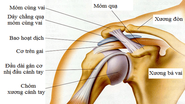
The shoulder joint is one of the major joints of the body, playing an important role in daily life, from delicate movements to vigorous activities such as sports, labor, and production.Shoulder joint is made up of many components:
- Bone around shoulder: humerus, shoulder blade, collarbone
- Joint: there are 03 main joints which are alveolar-brachial joint, sternum-clavicular joint, sacral joint shoulder – clavicle
- Cuff rotation: made up of shoulder cartilage, shoulder capsule and shoulder muscle.

Khớp vai giúp thực hiện nhiều động tác phục vụ đời sống và sinh hoạt hàng ngày
2. Some common shoulder joint diseases
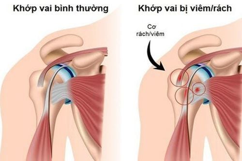
Some common basic diseases in the shoulder joint:Shoulder trauma: shoulder fracture, shoulder dislocation, tear - cuff damage..... Inflammation around the shoulder joint: due to degeneration, due to gout... .. Shoulder bursitis.

Viêm khớp là tình trạng thoái hóa phổ biến ở người lớn tuổi
3. X-ray technique of straight shoulder joint
3.1 Cases that the doctor appoints to take an x-ray of the shoulder joint When experiencing shoulder injuries due to causes such as: traffic accidents, labor accidents, accidents in daily life... causing pain shoulder joint pain Unexplained shoulder pain Shoulder joint pain accompanied by inability to move the arm Elderly shoulder pain 3.2 Procedure for taking X-ray of the shoulder joint Prepare the patient: Invite the patient to the imaging room, Explain the imaging process to the patient. Instruct the patient to undress to expose the area to be photographed and to remove any body jewelry, if any.
Bệnh nhân tháo bỏ trang sức hoặc đồ kim loại trước khi tiến hành chụp
Instruct the patient to stand or sit in front of the film stand, the patient should face the ball, the right hand does not need to be taken along the body, the right hand needs to be taken with the maximum arm shape perpendicular to the chest palm upturned hand. Adjust the patient to stand upright so that the back of the shoulder is close to the film. Set right and left marks. The x-ray ball is projected horizontally perpendicular to the film. The central ray is localized to the clavicle joint to be imaged. Instruct the patient to remain in the position. The distance of the film shadow is 1m to localize the x-ray beam. Check the buttons on the control cabinet, observe the patient through the glass, press the x-ray generator button. Invite the patient out of the waiting room. 3.3 Evaluation of the results After the clavicle was captured in the middle of the film, the scapula was separated from the ball of the suprahumeral head. Movies have contrast sharpness. clean film without scratches. Return the film, return the results to the x-rayer. Master, Doctor Nguyen Hong Hai graduated with a Master's degree in Diagnostic Imaging from Hanoi Medical University with strengths in diagnosing breast and thyroid cancer. Currently, Dr. Hai is working at the Department of Diagnostic Imaging, Vinmec Times City International Hospital.
Any questions that need to be answered by a specialist doctor as well as customers wishing to be examined and treated at Vinmec International General Hospital, you can contact Vinmec Health System nationwide or register online HERE.
LEARN MORE
Treatment methods for shoulder dislocation Rehabilitation exercises for periarthritis of the shoulder Partial shoulder dislocation surgery


