This is an automatically translated article.
The article was written by Dr. Bui Xuan Truong - General Internal Medicine Department - Vinmec Times City International HospitalLight is an entity in the natural cycle of day and night, helping us to distinguish things, from familiar people to everyday objects, trees, houses... Light also helps create energy. quality of life service. In medicine, light helps to create diagnostic and therapeutic devices, and one of those devices is the flexible gastrointestinal endoscope.
1. Some basic structural and functional features of the gastrointestinal tract
The basic structure of the alimentary canal wall consists of 4 layers, from the inside out including the mucosal layer Submucosal layer (submucosa) Muscular layer (inner sphincter and outer longitudinal muscle) Outer shell Tube The digestive system has the function of transporting and storing food, as well as the function of digesting and absorbing nutrients in food. Then, the excess food residue will be concentrated into feces, and then excreted through the anus. In the lining of the alimentary canal there are digestive glands. Depending on the specific structure and function of the alimentary canal (esophagus, stomach, small intestine, colon and rectum), the surface mucosal cells along with the digestive gland at a particular site may own characteristics. The submucosa is a connective tissue with blood vessels, nerves and lymphatic systems that nourish and transport toxins out.
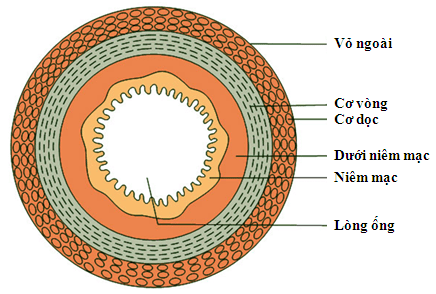
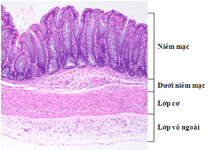
Hình ảnh mô tả các lớp của thành ống tiêu hóa
2. Why endoscopy can analyze the lining of the digestive tract
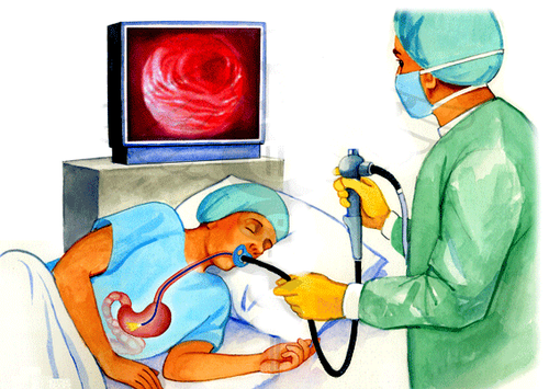
Flexible endoscopy is considered one of the outstanding technological inventions of the 20th century, allowing the widespread implementation of gastrointestinal endoscopy in the diagnosis and treatment of diseases. Currently, all new generation endoscopes are high resolution machines. The flexible gastrointestinal endoscope consists of the following main components:Flexible endoscope: Has a light transmission system and receives light signals, and there is a channel in the wire to insert instruments (bio-needle). equipment, needles, electrocautery forceps, hemostatic forceps, cutters, ligation knives, etc.) to conduct specimen collection for diagnostic work or to conduct treatment of a number of diseases (hemostatic injections, ligation of varicose veins, cutting polyps, cutting mucous membranes to treat early and very early cancers....). In addition, the bronchoscope also has a special small channel to inflate and pump water to clean the tip of the bronchoscope during the procedure. The system receives and processes image signals to produce real images during endoscopy. This system integrates with the inflatable and water pump system Screen The process of endoscopic treatment intervention may require additional equipment, such as: Electrocoagulation, booster water pump...
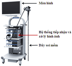
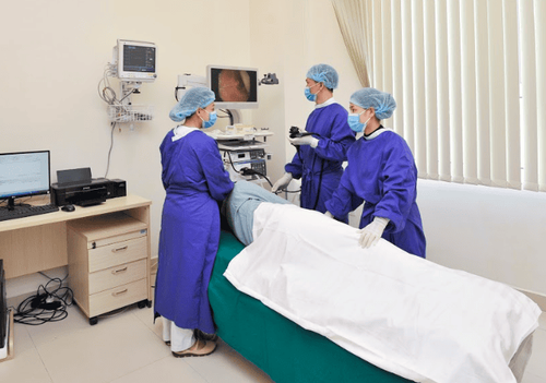
Mô tả hệ thống nội soi ống mềm Olympus CV-190 hiện đại hàng đầu thế giới, tại Bệnh viên Đa khoa Quốc tế Vinmec
During endoscopy, the digestive tube will be inflated and inflated for viewing. The light source coming out of the endoscope will interact with the wall layers of the digestive tract, especially the mucosa and submucosa.
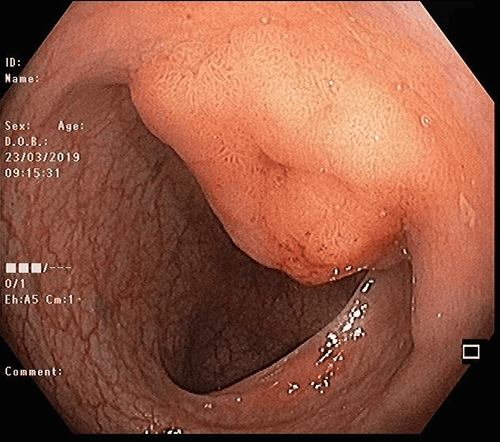
Natural light has many different wavelengths, so there will be a wavelength that penetrates the wall of the digestive tract deeper and has a shallower wavelength, with a wavelength that only interacts right at the mucosal surface layer. The signals of the reflected rays of light during the piercing process will be received and transmitted to the image center of the machine. Real images during endoscopy will be displayed on the machine screen. Cells in the body in general and cells lining the digestive tract in particular, when in a normal state, in an inflammatory state or transformed into cancer cells, will have different characteristics both in terms of the surface coating properties. surface, density of substances and water content inside the cell, cell nucleus size. In addition, there are changes in the blood supply to the submucosa of the gastrointestinal tract. These blood vessels are irregular in size and are located in different shallow and deep positions. Therefore, in each state of normal cells, inflammatory or cancerous cells will give characteristic features in morphology and color. Based on this change, the doctor through the image displayed on the endoscope screen will diagnose whether the patient has an inflammatory condition, is likely to have cancer or has cancer.
3. Why is NBI endoscopy of good diagnostic value?
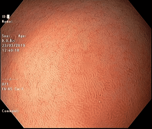
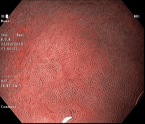
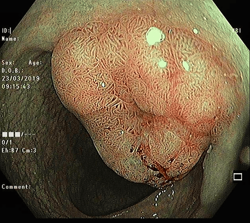
Different from conventional endoscopy using natural light, NBI (Narrow Band Imaging) endoscope system parallels two modes:Normal high-resolution endoscope mode using natural light with step wave oscillation between 400-700 nm Narrow band imaging (NBI: Narrow Band Imaging) mode uses only light with 415 nm wavelength and 540 nm wavelength
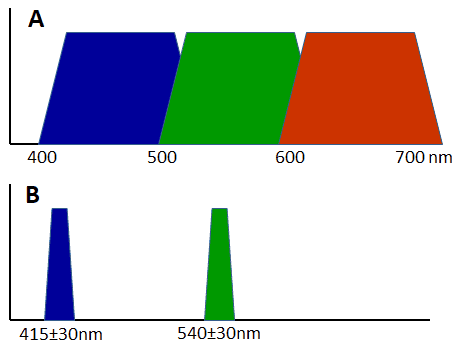
Hình ảnh mô tả sự khác biệt trong áp dụng sử dụng bước sóng ánh sánh giữa (A): Nội soi độ phân giải cao thông thường và (B): Nội soi với dải tần ánh sáng hẹp NBI (Narrow Band Imaging)
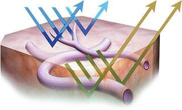
Hình ảnh mô tả hai bước sóng ánh sáng áp dụng trong nội soi NBI cho phép phân tích tốt hơn bề mặt niêm mạc và mạng lưới mao mạch nông
(A)
(B)
(A)
(B)
Vinmec International General Hospital is equipped with a high-resolution flexible gastrointestinal endoscope - the world's leading high-quality NBI endoscope of Olympus Company (Japan). Therefore, at Vinmec Hospital, there are many patients with high risk of gastrointestinal cancer or gastrointestinal (esophageal - stomach - colorectal) cancer in early stage, very early stage being treated. Successful endoscopic mucosal resection – submucosal dissection.
Please dial HOTLINE for more information or register for an appointment HERE. Download MyVinmec app to make appointments faster and to manage your bookings easily.


