This is an automatically translated article.
Esophageal perforation is a serious medical problem, with a high risk of death if not detected and treated promptly. Currently, the diagnosis of esophageal perforation by X-ray technique is the most optimal method chosen by doctors.
1. Causes of esophageal perforation
The esophagus is the first part of the digestive tract, about 25cm long. The function of the esophagus is to carry food and water from the mouth to the stomach. The position of the esophagus is parallel to the trachea and to the left of the heart, in front of the spine. This is an organ with many connective tissues and blood vessels passing through to cover and protect.Esophageal perforation is uncommon but it is a very serious medical condition that is life-threatening if not detected early and treated within 24 hours. The esophagus is divided into 3 segments, corresponding to the neck, thoracic and abdominal regions. Esophageal perforation can occur at any site.
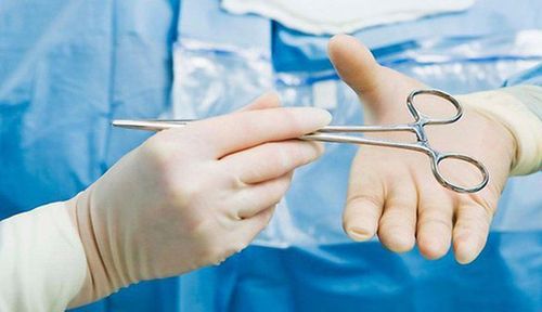
A common cause of esophageal perforation is the use of medical instruments in medical procedures that are not technically correct. In addition, the following less common causes can also cause esophageal perforation:
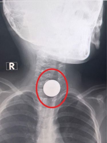
Tumor in the throat; sore throat caused by gastroesophageal reflux disease (GERD); Accidentally swallowing foreign objects, acids or chemicals; Injuries, accidents in the neck; Violent vomiting.
2. Signs of esophageal perforation patients should know
The first signs of esophageal perforation are pain at the site of the perforation, chest tightness, and difficulty swallowing food. Besides, the patient may experience other symptoms such as:
Heart palpitations; Rapidly breath; Low blood pressure ; Fever; Chills; Vomiting, possibly vomiting blood; If the perforation is in the neck area, the patient will feel pain or stiffness in the neck.

Dấu hiệu đầu tiên của thủng thực quản là đau ở vị trí thủng, tức ngực và khó nuốt thức ăn hơn.
3. Medical techniques to help diagnose esophageal perforation
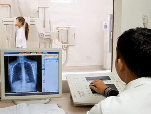
To diagnose esophageal perforation, the doctor will ask the patient to perform tests to evaluate the condition, see if there is a leak of fluid out of the esophagus or into the lungs. Esophageal X-ray technique is the most optimal method of choice for the diagnosis of esophageal perforation.
When performing an esophageal X-ray, the patient must fast, not drink water, do not bring jewelry or metal objects into the imaging room.
The doctor will guide the patient to lie down or stand depending on the purpose of the survey. During the scan, the patient takes a mouthful of contrast medium and swallows according to the doctor's instructions. During the scan, the patient may need to hold their breath for a certain amount of time for the image to be clear. Esophageal X-ray procedure takes about 30 minutes.
Considered as an optimal, low-cost and quick-effective imaging method, esophageal X-ray helps doctors make early diagnoses related to esophageal perforation, thereby having a method. fastest treatment.
In addition to esophageal radiography, chest CT can detect gas and fluid but cannot localize the perforation site. The endoscopic method can bypass the small perforation.
4. Effective treatment of esophageal perforation
Treatment of esophageal perforation must be carried out as soon as possible, to prevent infection from occurring. Patients have a high chance of survival if they are successfully treated within 24 hours of diagnosis. Depending on the condition of the esophageal perforation, the doctor will recommend the most appropriate treatment.
First, the doctor will drain all the fluid out, the patient is prescribed antibiotics to prevent or treat infection. During the treatment of esophageal perforation, the patient is not allowed to eat or drink, nutrients are given through a feeding tube, and drugs and fluids are given intravenously.
If the perforations are small, the fluid inside the esophagus does not back up and leak into the lungs, then no surgical intervention is needed, the perforations will heal on their own.
However, most cases of esophageal perforation are indicated for surgery. If the hole is small, the doctor will sew it up, but if the hole is too large, it is necessary to cut part of the esophagus and reconnect it to the stomach.
Esophageal perforation, if not detected and treated early, can cause many life-threatening complications such as: pneumonia, blood infection, empyema, acute respiratory failure... Therefore, If signs of esophageal perforation are suspected, the patient should quickly go to a reputable medical facility for examination, diagnosis and treatment.
Currently, Vinmec International General Hospital is one of the top quality hospitals in the country, trusted by a large number of patients for medical examination and treatment. Not only the physical system, modern equipment: 6 ultrasound rooms; 4 DR X-ray rooms (1 full-axis, 1 intensifier, 1 synthesizer and 1 mammogram); 2 portable X-ray machines DR; 2 multi-row computer tomography rooms with receivers (1 128 rows and 1 16 rows); 2 rooms for magnetic resonance imaging (1 machine 3 Tesla and 1 machine 1.5 Tesla); 1 room for 2 levels of interventional angiography and 1 room to measure bone mineral density.... Vinmec is also the place where a team of experienced doctors and nurses will gather a lot of support in diagnosis and detection. early signs of abnormality in the patient's body. In particular, with a space designed according to 5-star hotel standards, Vinmec ensures to bring the patient the most comfort, friendliness and peace of mind.
Please dial HOTLINE for more information or register for an appointment HERE. Download MyVinmec app to make appointments faster and to manage your bookings easily.


