This is an automatically translated article.
The article was professionally consulted by Specialist Doctor II Nguyen Quoc Viet - Department of Medical Examination & Internal Medicine - Vinmec Danang International General Hospital. The doctor has more than 20 years of experience in the examination and treatment of cardiovascular diseases and Cardiovascular Interventions (Including angiography, dilation, stenting of coronary arteries, renal arteries...), placing temporary pacemakers , forever...The normal ST segment lies on the isoelectric line, with very little difference up or down the isoelectric line. The image of ST-segment elevation or ST-segment depression on electrocardiogram results is valuable in the diagnosis of some cardiovascular diseases.
1. What is an ST segment?
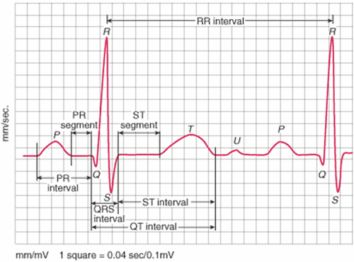
On the electrocardiogram, the ST segment does not consist of a single wave but is a straight line from the end of the QRS to the beginning of the T wave. The ST segment represents the end of depolarization to the beginning of repolarization. pole.ST segment is normal when 75% of ST segment is above the isoelectric line, if elevation is not more than 0.1 mV and depression is not more than 0.05 mV. In general, the ST segment does not curve but goes straight and smoothly continues into the T, nor does it ever go downhill, but only sideways or slightly uphill.
ST segment duration is relatively long and depends on heart rate. However, ST segment duration is of little clinical use. On the contrary, the shape and position of the ST segment relative to the isoelectric line is a new feature that is noticeable and meaningful in diagnosis.

2. ST segment elevation in what cases?
ST segment elevation is defined as ST elevation in at least two consecutive leads and the following conditions are met:> 0.1 mV in the periphery and V4-V6 > 0.2 mV in V1-V3 ST segment elevation in the following conditions:
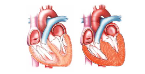
Acute ST-elevation myocardial infarction (acute STEMI): can produce short convex or concave ST elevation or indirect morphology, often with depression in the contralateral leads. Coronary vasospasm: causes ST-segment elevation similar to acute STEMI, but the ECG changes are transient, and the ST segment usually returns to normal when pain subsides or vasodilators are used. Electrocardiographic changes may not be distinguished. Benign early repolarization: causes mild ST elevation with T waves, often with a V-shaped notch of the J point. This is a normal variant, common in children and healthy patients. Left bundle branch block: Produces high ST with vertical T waves in leads with negative QRS (dominant S waves), while negative ST inversion reverses T waves in leads with positive QRS complexes. Acute pericarditis: Causes ST elevation but depression with PR depression in multiple leads. Commonly seen in I, II, III, aVF, aVL and V2-V6. In leads aVR and V1 there is reciprocal ST depression. Left ventricular hypertrophy: Causes an appearance similar to left bundle branch block, high ST in leads with deep S waves, usually V1-V3, and deep ST/T inversion in leads with high R (I, aVL) , V5-V6).
Trắc nghiệm: Huyết áp của bạn có đang thực sự tốt?
Huyết áp cao hay thấp đều ảnh hưởng đến tình trạng sức khỏe con người. Để biết tình trạng huyết áp của bạn có thực sự tốt không, hãy làm bài trắc nghiệm sau đây để đánh giá.
3. In what cases does ST segment depression appear?
ST-segment depression >0.5mm has diagnostic significance. There are many types of ST segment depression such as: oblique depression, horizontal depression, concave depression, descending depression, cup-shaped concave depression.ST segment depression is common in the following conditions:
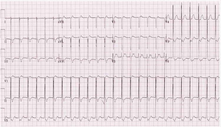
Myocardial ischemia: ST depression may be present in some leads, most commonly in the left precordial leads V4-V6 with wide ST depression and ST elevation in aVR in left main coronary artery occlusion. The corresponding ST elevation may be difficult to see but should be sought. Posterior MI: acute ST-segment elevation myocardial infarction causes symmetrical St depression in the anterior lead in V1-V3, along with a dominant R wave and a vertical T wave. ST elevation in lead V7-V9. Digoxin toxicity causes ST depression with a “hammock” appearance. Hypokalemia: causes wide ST-segment depression, flat/inverted T waves, prominent U waves, and a prolonged QU interval. Right ventricular hypertrophy: causes ST depression, T wave inversion in the right precordial lead V1-V3. Right bundle branch block: ST depression and T wave inversion in V1-V3. Supraventricular tachycardia: usually causes wide ST elevation and depression, prominent in the left precordial leads V4-V6.

To protect heart health in general and detect early signs of myocardial infarction and stroke, customers can sign up for the Cardiovascular Screening Package - Basic Cardiovascular Examination of Vinmec International General Hospital. The examination package helps to detect cardiovascular problems at the earliest through tests and modern imaging methods. The package is for all ages, genders and is especially essential for people with risk factors for cardiovascular disease.
Please dial HOTLINE for more information or register for an appointment HERE. Download MyVinmec app to make appointments faster and to manage your bookings easily.


