This is an automatically translated article.
Posterior urethral valve is a congenital disease that occurs due to a disorder in the formation of the urogenital tubules of children during the fetal period. The frequency of this pathology in boys ranges from 1/5000 to 1/25,000.
1. What is the posterior urethral valve?
In boys, the urethra is divided into two parts: the anterior urethra and the posterior urethra (respectively the inner spongy part of the penis and the outer part of the spongy object).
Posterior urethral valve is a condition in which a diaphragm appears in the posterior urethra, making it difficult for urine to flow and can flow back into the bladder or even back into the ureters and kidneys. The stagnation and backflow of urine will cause extremely dangerous complications for children.
2. Signs of urethral valve
Posterior urethral valve occurs in young children and infants, so the diagnosis and treatment will be somewhat more difficult.
Children with urethral valve often have symptoms of crying because they cannot urinate, sometimes accompanied by fever, shortness of breath, and abdominal distension. Children often have slow growth and anemia. Examination revealed a bladder bridge, possibly enlarged kidneys. For older children, the signs are more obvious such as: Difficulty urinating, urinary incontinence, frequent urination...
Trẻ đi tiểu nhiều lần có thể là dấu hiệu mắc bệnh van niệu đạo sau
Due to not being able to urinate completely, the urge to urinate a lot also causes increased pressure on the abdomen. This can cause some diseases such as rectal prolapse, bladder fistula or hernia masses...
To detect the posterior urethral valve, pregnant women should have regular ultrasound scans during pregnancy. to track. Posterior urethral valve disease can be diagnosed prenatally around the 12th week of pregnancy.
3. Is the urethral valve dangerous?
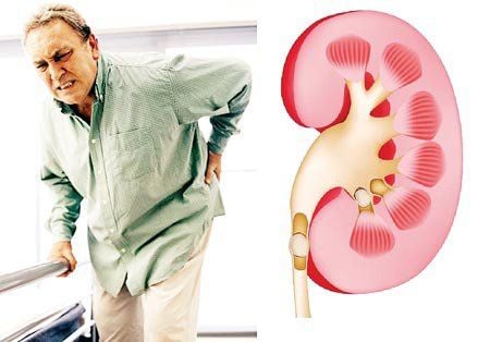
Obstruction of the urethral outlet will cause urine stagnation, causing the bladder lumen and the first part of the posterior urethra to dilate. The stagnation of urine will lead to infection, the consequences of a long-term inflammatory process will cause the bladder wall and two ureteral openings to be deformed.
Due to the influence of increased pressure occurring in the bladder lumen, leading to reflux of urine through the 2 ureteral openings. Sometimes, reflux can continue even after the urethral valve has been removed, which is due to a malformation of the triangle of the bladder. Posterior urethral valve is also the leading cause of obstruction that can cause renal failure.
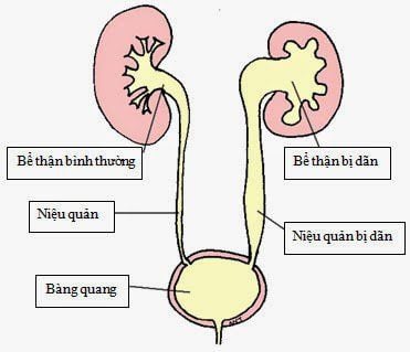
If the vesicoureteral reflux is prolonged, it will lead to dilation of the ureters, dilatation of the calyces - pyelonephritis and diseases that are difficult to recover even after removing the urethral valve. Therefore, it is necessary to take the child with a urethral valve after going to the doctor and remove it as soon as possible before bad complications occur.
Bệnh không được điều trị kịp thời dễ dẫn tới giãn đài bể thận
4. Diagnosis of posterior urethral valve
4.1 Prenatal diagnosis The posterior urethral valve can be diagnosed during pregnancy. That is around the 12th week, when the fetus begins to be able to filter urine. The first signs of urethral valve disease are followed by pyelonephritis and lack of amniotic fluid.
4.2 Clinical and laboratory diagnosis 4.2.1 Neonates In neonates, symptoms are quite severe due to their young age. Systemic symptoms can include hypothermia, anemia, dehydration, jaundice... Respiratory depression and peritoneal effusion are also symptoms but are less noticeable. Meanwhile, the urinary tract symptoms are too vague.
4.2.2 Infancy At this age, urinary tract symptoms are more obvious. Children have symptoms such as large kidneys, difficulty urinating, urinary leakage, bladder bridges...
4.2.3 Older children The signs in older children are very clear, but a lot of straining to urinate can also cause other diseases. for children. In general, the symptoms are quite specific to the following urethral valve disease, including:
Difficulty urinating Leaky urine Bladder bridge Urinary tract infection To know the exact condition, the doctor may order additional tests such as abdominal ultrasound, retrograde cystography when urinating, urinary tract scan, nephrograph. Some kidney function tests such as blood and urine tests, urinary tract infection or blood infection may also be performed.

Chẩn đoán van niệu đạo sau ở trẻ bằng hình ảnh
4.3 Treatment of posterior urethral valve Depending on the severity and condition of the patient, doctors will have an appropriate treatment plan.
4.3.1 Neonates and infants In neonates and infants, the disease is usually advanced by the time it is present and causes severe systemic symptoms. Therefore, it is necessary to focus on treating the whole body first and then specializing in treatment.
Systemic treatment includes:
Cardiovascular and respiratory resuscitation, Acid-base balance, Use of antibiotics to fight infection, Rehydration of fluids and electrolytes Improve the patient's condition After recovering, doctors The doctor has just started going into specialized treatment. Depending on each case, the treatment options may vary, but generally include:
Urinary catheterization, bladder drainage Bringing the ureter to the skin If the health condition is good, endoscopic urethral valve resection can be performed 4.3.2 Older children In older children, the general condition is usually stable and the urethral diameter is also relatively large compared to neonates and infants. Therefore, in the case of older children with posterior urethral valves, the valve will be cut through urethroscopy. The valve will be cut with an electric knife. After cutting, if the test is unsatisfactory or still obstructs the urinary tract, it is necessary to do the valve surgery for the second time. For treatment at Vinmec International General Hospital, you can contact Vinmec Health System nationwide or register online HERE.


