This is an automatically translated article.
The article was professionally consulted with the Radiologist, Department of Diagnostic Imaging - Vinmec Central Park International General Hospital.The birth of ultrasound marked the development of world medicine. Because ultrasound will help doctors diagnose diseases deep inside the body that no previous method could do. Among them, the superiority and accuracy of echocardiography must be mentioned.
1. What is ultrasound?
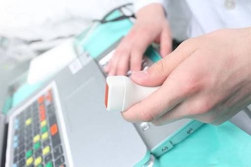
Medical ultrasound is a method of diagnosing diseases using transducers that emit ultrasound waves. High frequency ultrasound waves have the function of probing the parts below the skin and then reflecting back through medical images. Thanks to the development of mechanical devices, ultrasound images are becoming clearer and more realistic, helping doctors to easily diagnose and detect diseases.
2. Common types of ultrasound
Ultrasound has different techniques for each ultrasound object. But the most popular are still the following techniques:
2.1 3D ultrasound 3D ultrasound is one of the most applied medical ultrasound techniques today. 3D ultrasound is often used to diagnose many diseases, in which it is often used in antenatal care. Through the tissue, the ultrasound probe will reflect back and give a clear image with 3 spatial dimensions.
2.2 Doppler Ultrasound Doppler ultrasound is capable of measuring the flow of movement in blood vessels. This method is most commonly applied to fetal and cardiovascular ultrasound. Thanks to Doppler ultrasound, doctors can early detect fetal abnormalities as well as the nutritional status of the baby in the womb. In particular, the Doppler method can detect fetal heart defects very accurately that no other method can do.

Siêu âm Doppler thường được sử dụng trong siêu âm thai nhi và siêu âm tim mạch
Doppler ultrasound is a safe method for both mother and baby, especially for pregnant women who are in the last 3 months of pregnancy, they should go for Doppler ultrasound to be able to know the most accurately about the baby's condition before at birth.
There are two main types of Doppler ultrasound: pulsed ultrasound and continuous Doppler ultrasound.
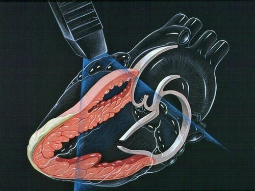
2.3 Echocardiography Echocardiography is a technique that uses ultrasound waves to study the structure and diagnose heart diseases. The ultrasound signals will reflect the contraction of the working heart through clear and accurate images. Not only that, doctors can also assess the position, posture, size of the heart chambers, be able to detect holes in the septum in the heart, malformations or tumors in the heart, pericardial fluid. heart.
There are common types of echocardiography such as:
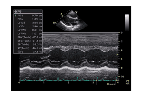
TM echocardiography; 2D echocardiography; 3D echocardiography; 4D echocardiography ; Doppler echocardiography. 2.4 Ultrasound therapy This method is commonly applied in physical therapy, helping to treat problems such as pain or damage to the skin or soft tissues.
Therapeutic ultrasound has thermal and mechanical effects on human tissue and skin. By using the appropriate frequency, the tissues will easily absorb the sound wave energy. This method can help relieve pain or regenerate damaged tissues inside the human body.
Thus, echocardiography is only one method in general ultrasound. Therefore, echocardiography is only a small part of ultrasound, only affecting the cardiovascular system. Compared with ultrasound, echocardiography is not as technical as it is, but it is accurate to the heart organs, which if only using conventional ultrasound, will not detect.

Kỹ thuật siêu âm trị liệu
3. Echocardiography can detect what diseases?
With ultrasound, the doctor can see the structure of the heart and check for abnormalities when the heart is working. In particular, it will help doctors understand:
How the heart works, contracts; Heart size and shape; Size and pumping motion of the heart walls; The pumping power of the heart; whether the heart valves are working properly or not; Whether the heart valve is narrow or not; Is there blood regurgitation through the heart valves; Is there a tumor, an inflammatory mass around the heart valves, heart muscle, or blood vessels? Thereby, based on the results that the ultrasound shows, the doctor can diagnose diseases related to the heart:
Problems with the large blood vessels entering and leaving the heart; Problems with the heart muscle, inner and outer membranes of the heart; Heart valve diseases; Abnormal holes between the chambers of the heart; Blood clots in the heart chambers.
4. When is an echocardiogram needed?
The following signs are manifestations of some dangerous diseases, the patient should go to the medical center immediately:
The patient has unusual signs and symptoms such as chest tightness, shortness of breath, angina ;

Khi người bệnh có triệu chứng đau ngực cần được siêu âm tim
Heart valves are damaged; Check the functioning of the heart; For people who are assigned to have heart surgery, echocardiography for doctors to know the parameters; Echocardiography to understand the width of the orifice, whether the valve is closed or open; To detect fetal malformations; Monitoring of complications of the disease on coronary artery disease
5. What is the difference between echocardiography and conventional echocardiography?
When doing ultrasound, the patient does not need any special preparation before performing the ultrasound. During the ultrasound, the part that is technically manipulated is the heart part.
Echocardiography is carried out technically by 3 methods: exercise ultrasound, Doppler ultrasound, transesophageal ultrasound. Each ultrasound technique has different functions and uses.
Stress ultrasound: When the doctor conducts an electrocardiogram, or gives the patient medication that can make the heart beat faster and stronger. Doppler ultrasound is indicated to measure blood flow velocity at locations in the heart chambers. Transesophageal ultrasound: The patient must swallow a transducer with a thin fiber optic cable connected to the ultrasound machine.

Siêu âm tim cần được thực hiện bởi các bác sĩ chuyên khoa
6. Possible side effects or complications of echocardiography
Unlike conventional ultrasound, echocardiography will cause some side effects. Even so, patients should not worry, as these are very normal phenomena.
Echocardiography is a modern method of diagnosis and cardiovascular examination, giving clear images.
Furthermore, conventional transthoracic echocardiography is painless and uncomplicated. Some side effects may be encountered such as:
In the case of having electrodes to monitor the electrocardiogram at the same time, the patient will feel discomfort when the doctor removes the adhesive tape attached to the electrodes on the chest. Throat pain for several hours if transesophageal echocardiography, rarely the ultrasound tube scratched the inner throat. During the ultrasound, you may experience breathing problems due to the sedation or the amount of oxygen you breathe. Exercise or medication during stress echocardiography can cause transient arrhythmias, not echocardiography. The heart is an organ directly related to health. Therefore, when there are signs of heart disease, patients need to seek medical attention for ultrasound, early detection of disease for effective treatment.
To protect cardiovascular health in general and detect early signs of cardiovascular disease, customers can sign up for Cardiovascular Screening Package - Basic Cardiovascular Examination of Vinmec International General Hospital. The examination package helps to detect cardiovascular problems at the earliest through tests and modern imaging methods. The package is for all ages, genders and is especially essential for people with risk factors for cardiovascular disease.
Please dial HOTLINE for more information or register for an appointment HERE. Download MyVinmec app to make appointments faster and to manage your bookings easily.