This is an automatically translated article.
The article is professionally consulted by the Doctor of Cardiology Interventional Unit, Vinmec Times City International General Hospital.Doppler echocardiography is one of the common imaging techniques used in the diagnosis, monitoring, and observation of heart movement, function of myocardium, heart valves and many other cardiovascular diseases. . The Doppler echocardiography procedure is usually quite quick, and can be done in the ultrasound room or emergency ultrasound at the bed.
1. What is echocardiography?
Echocardiography is a method that uses high-frequency ultrasound waves to view and obtain images of the heart, movements or structures related to the heart. Based on the above images, the doctor can make an initial assessment of the shape, size, as well as abnormal features of the heart, heart valves.
Siêu âm tim là phương pháp sử dụng sóng siêu âm từ đầu dò giúp phát hiện các dấu hiệu bất thường của tim
2. Benefits of Echocardiography
During ultrasound, the doctor will use transducers with waves, moving around the patient's chest or esophagus. The ultrasound process helps doctors easily observe the following problems:
Shape, size, activity, contraction of the heart. Size and pumping of the valves and walls of the heart. The pumping power of the heart and the volume it pumps blood to other organs of the body. This pumping power is calculated in minutes and averaged. Check the heartbeat. Detect abnormalities or damage to the heart such as open heart valves, wide or narrow heart valves, abnormal blood clots in the heart, problems with the outer lining of the heart. Detecting abnormal tumors of unknown cause, causing compression and pain in the chest.

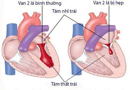
Hình ảnh siêu âm tim Doppler màu hở van hai lá
Echocardiography is an effective solution to help doctors diagnose as well as easily detect heart-related diseases such as heart valve regurgitation, heart failure, myocardial infarction, myocarditis,... This helps patients have time to prepare psychologically and quickly receive timely treatment to help ensure quality of health before worsening conditions occur.
3. When should the patient perform echocardiography?
You should perform this ultrasound method when you see the body has abnormal signs related to the heart. There are even cases where the doctor will appoint the patient to conduct an ultrasound to get an accurate conclusion based on specific images of the structure and functioning of the heart. Specific cases can be mentioned as follows:
The examiner has prolonged chest pain, throbbing chest pain, difficulty breathing. The patient has signs related to heart disease such as: dizziness, anemia, chest pain, bad breath of unknown cause, ... The patient was diagnosed with abnormal signs. heart problems such as fibrillation, irregular heartbeat, and instability. Patients with a history of cardiovascular disease, or a family history of heart disease. The test results were abnormal and related to cardiovascular problems.

Khi xuất các dấu hiệu như đau tức ngực kéo dài không rõ nguyên nhân, khó thở là lúc bạn nên tiến hành siêu âm tim và thăm khám tim mạch
Usually, patients only go to the doctor when they notice abnormal signs. This is the reason why the disease is discovered when it has entered a serious stage, reducing the ability to treat or endangering the patient's life, and expensive in treatment costs. Therefore, each person should actively perform echocardiography periodically to ensure the quality of health, as well as promptly treat if abnormal heart diseases are detected.
4. Cardiac Doppler echocardiography
4.1. Prepare before the ultrasound
Unlike other forms of ultrasound, you can still eat and drink completely normal before the ultrasound. However, in some cases where a transesophageal or stress ultrasound is required, the doctor will actively ask you to fast for a few hours before the ultrasound. The transesophageal ultrasound will require medication. sedation, so you should have a loved one with you so that it is safest to go home after the ultrasound. In addition, you should prepare psychologically during the ultrasound. This is also an important factor for the most accurate images and results of cardiovascular status.

Người bệnh có thể cần sử dụng thuốc an thần khi siêu âm qua thực quản
4.2. Ultrasound process
Unlike other forms of ultrasound, you can still eat and drink completely normal before the ultrasound. However, in some cases where a transesophageal or stress ultrasound is required, your doctor will actively ask you to fast for a few hours before the ultrasound. The transesophageal ultrasound will require medication. sedation, so you should have a loved one with you so that it is safest to go home after the ultrasound. In addition, you should prepare psychologically during the ultrasound. This is also an important factor for the most accurate images and results of cardiovascular status.
4.2. Ultrasound process
Usually, the ultrasound will take about 30 minutes to 1 hour. However, this period may be less or longer depending on the associated cardiovascular damage.
The ultrasonic process is different with each form. Specifically as follows:
Echocardiography through the chest area: the doctor applies a gel to the chest to increase the wave conduction. The transducer is moved continuously over the chest, and images of the heart are captured on a monitor. When abnormal signs are detected, the doctor will take relevant images to diagnose cardiovascular diseases. With a transesophageal echocardiogram, you will be sedated with an anesthetic inhaler or sedated. The endoscope is then inserted into your throat. The doctor will push the tube deep into the esophagus and take pictures of the right and left chambers of the heart. At each position, the probe is held for 2 - 3 minutes.
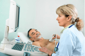
Siêu âm quanh vùng ngực giúp ghi lại những hình ảnh về cấu trúc và hoạt động của tim . Đây là cơ sở giúp chẩn đoán và phát hiện các bệnh lý bất thường liên quan đến tim mạch
4.3. After ultrasound
After the ultrasound process, you will receive diagnostic examination results based on the captured and captured images. If cardiac abnormalities are found, you may be recommended to do some more tests, explore to give the final results or consult and give treatment directions if immediately detect specific pathology.
Doppler echocardiography is used to evaluate many different medical conditions. Follow-up will depend on the final outcome of this procedure. In summary, the follow-up process after echocardiography is mainly to treat abnormalities or cardiovascular diseases of the patient.
To protect heart health in general and detect early signs of myocardial infarction and stroke, customers can sign up for Cardiovascular Screening Package - Basic Cardiovascular Examination of Vinmec International General Hospital . The examination package helps to detect cardiovascular problems at the earliest through tests and modern imaging methods. The package is for all ages, genders and is especially essential for people with risk factors for cardiovascular disease.
Please dial HOTLINE for more information or register for an appointment HERE. Download MyVinmec app to make appointments faster and to manage your bookings easily.
LEARN MORE
Evaluation of pulmonary artery pressure by echocardiography Doppler echocardiography Application of echocardiography Doppler echocardiography procedure