This is an automatically translated article.
Papillary edema is an important fundus symptom for endocarditis, with multiple causes including orbital, ocular, intracranial, and systemic.
1. What is thorns?
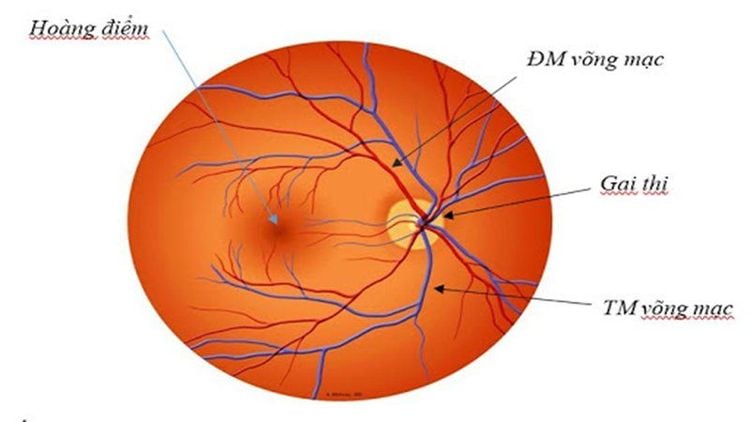
The optic nerve is where the optic nerve enters the eyeball. When looking at the fundus, you will see that the optic nerve ending is the optic nerve. The central retinal vein and artery also pass through this site to enter the eyeball.
The optic spine is deviated to the nose and is home to the axons of 1-1.2 million retinal ganglion cells, which are unmyelinated. The normal optic nerve is round or oval in shape, with an average horizontal diameter of 1.7 - 1.8 mm, an average longitudinal diameter of 1.85 - 1.95 mm. In the center of the posterior pole of the retina, about 4 mm from the optic disc to the temple, there is a small hole with many small cones and is the area that gives the best vision called the macula.

Vị trí của gai thị trong nhãn cầu
2. What is papilledema?
Papilledema is swelling and blurring of the optic disc margin, which occurs due to increased intracranial pressure or nerve compression injury. Papilledema is an important ophthalmic symptom for endocarditis. Optic papilledema results from obstruction of cytoplasmic outflow in the axons of the optic nerve, leading to axonal edema of the optic disc.
Papilledema is associated with other signs of optic nerve dysfunction, eg, decreased visual acuity, visual field loss, abnormal afferent pupillary reflex. The most common causes of papilledema are physiological blind spot enlargement, tubular visual field, and subnasal field loss.
Stages of papilledema :
Early stage: difficult to define, signs are papilledema, papillae margins are slightly blurred in the temporal part, papillae margins are slightly raised and veins are slightly dilated. Complete development stage: optic disc protrudes fungal form and loses physiological concave spine. The layers of nerve fibers emerge as light brown rays, the papilledema margin is erased, lying in a layer of diffuse edema fluid. The major vascular roots are obscured and the veins are twisted and dilated. Hemorrhage on the spine around the spine. Chronic phase: papilledema lasts for months and bleeding, discharge gradually dissolves. The arteries are narrowed, the veins are less tense, and the protrusion of the optic disc is also reduced. Central visual field begins to decrease, chromatic aberration, peripheral vision defect. The stage of atrophy of spines: the spines become more and more discolored, white, the edges are jagged, and the shadow is lost. The blood vessels on the spines disappear, the retina shrinks. Loss of vision is severe and can lead to blindness.

Phù gai thị dẫn đến rối loạn các chức năng thần kinh thị giác
3. Causes of papilledema
There are many causes of papilledema such as ocular, intracranial, orbital and systemic.
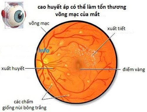
Eyeball: sudden drop of the eyeball several times due to a puncture wound to the eyeball. Occlusion of the central retinal vein, or glaucoma in glaucoma,... Orbit: pressure on the optic nerve in the orbit can lead to papilledema. Some diseases such as orbital abscess, orbital tumor, protrusion caused by endocrine disease basedow,... Intracranial: intracranial causes such as primary intracranial tumor and metastatic orbital tumor, sylvius aqueduct, epidural and subdural hematomas due to trauma, subarachnoid hemorrhage, severe headache and possibly anterior retinal hemorrhage, cerebral arteriovenous malformation, encephalitis,... Whole body : causes whole body such as hypertension, pregnancy toxicity, white blood cell,...
4. Symptoms of papilledema
Papilledema usually occurs in both eyes and enlarges the blind spot without causing loss of vision. Specific manifestations of papilledema include:
Headache, double vision, nausea and vomiting, rarely decreased vision. Visual field defects and decreased central vision if papilledema is chronic. Swelling and congestion of the optic disc on both sides. Retinal hemorrhage in the papilla or around the spine, dilated retinal veins, white cotton dots, zigzag, pupillary reflex, normal chromatic acuity, and widening physiological blind spot.

Đau đầu có thể là một trong những triệu chứng của phù gai thị
For chronic papilledema, special monitoring of the visual field and optic nerve sheath is required. Chronic papilledema will progress to papillae atrophy, hemorrhage, and white cotton spots that will disappear. The blood vessels around the spine will thicken and narrow, around the optic disc will form sphincter vascular shunts - the ciliary body. And eventually lead to chromosomal damage, central vision and visual field defects.
5. Treatment of papilledema
Treatment for papilledema will depend on the cause. However, diagnosing the cause is extremely complicated. Therefore, first of all, it is necessary to solve the circulatory and nutritional disorders in the nerves, fight infections, and fight inflammation.
To summarize, papilledema is the swelling and dilation of the optic disc due to increased intracranial pressure or damage to the optic nerve. If not treated early enough, vision loss can lead to blindness. Therefore, when having symptoms of headache, double vision, etc., it is necessary to immediately go to a medical facility and have regular eye exams, for early detection and timely treatment measures.
To visit and treat at Vinmec, please go directly to Vinmec health system or register online HERE.


