This is an automatically translated article.
The article was professionally consulted MSc Vu Van Quan - Gastroenterologist - Department of General Surgery & Anesthesiology - Vinmec Hai Phong International General HospitalThe mesentery is a part of the digestive system and performs many important functions in the human body. This is a newly discovered organ, bringing the total number of parts in the human body to 79.
1. Definition of mesentery
The mesentery or mesentery becomes a new organ in the body. Through anatomical and structural studies of the mesentery, it is shown that it is a monolithic part and performs many important functions.
The mesentery is the part of the peritoneum, the lining of the abdominal cavity, that attaches the intestine to the abdominal wall and holds the intestine in place. Some argue that the mesentery is a separate organ, as important as the other constituent parts of the body.
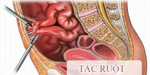
The mesentery connects the intestine to the abdominal cavity. It also helps bowel movements to a certain extent in the abdomen. Without the mesentery, the intestines are likely to frequently twist and turn and cause obstruction.
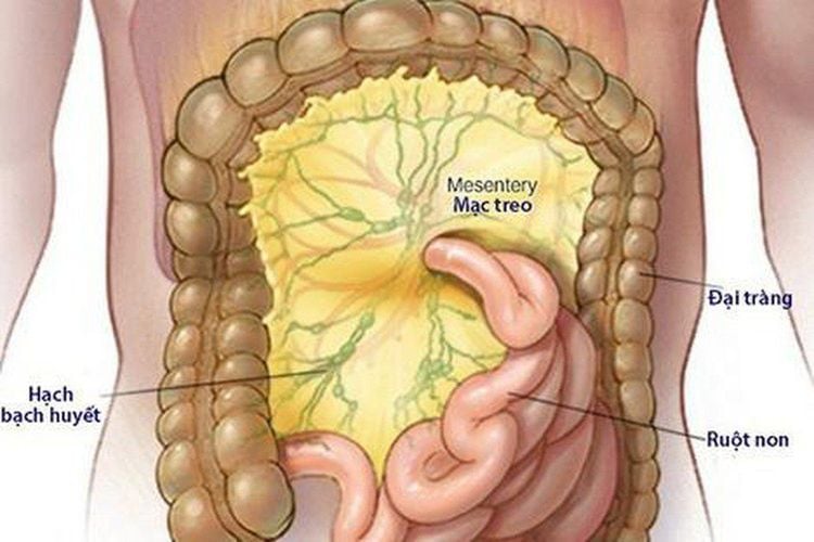
Mạc treo trong ổ bụng gắn ruột với khoang bụng
2. Types of mesentery
The mesentery is the medial peritoneal (including two leaflets) located between the peritoneum covering some segments of the intestine and the posterior abdominal peritoneum, which is the means of hanging these intestinal segments to the abdominal wall and creating the passageway for vessels. and nerves go to the intestinal segments. There are four mesenteries: the mesentery of the small intestine, the mesentery of the transverse colon, the mesentery of the sigmoid colon, and the mesentery of the appendix.The mesentery of the small intestine resembles a corrugated paper fan. It has a root about 15 cm long that attaches to the posterior abdominal wall along an oblique line from the left flank of the body of the second lumbar vertebra to the right sacroiliac joint, crossing in turn: transverse part of duodenum, abdominal aorta, inferior vena cava, right ureter, and right lumbosacral muscle. The average width from the mesenteric root to its intestinal margin is about 20 cm, but it is larger at the midpoint. Structures located between the leaflets of the mesenteric small intestine include: the jejunal and ileal branches of the superior mesenteric vessels, nerves, chyme vessels, and lymph nodes, along with a variable amount of fat. .
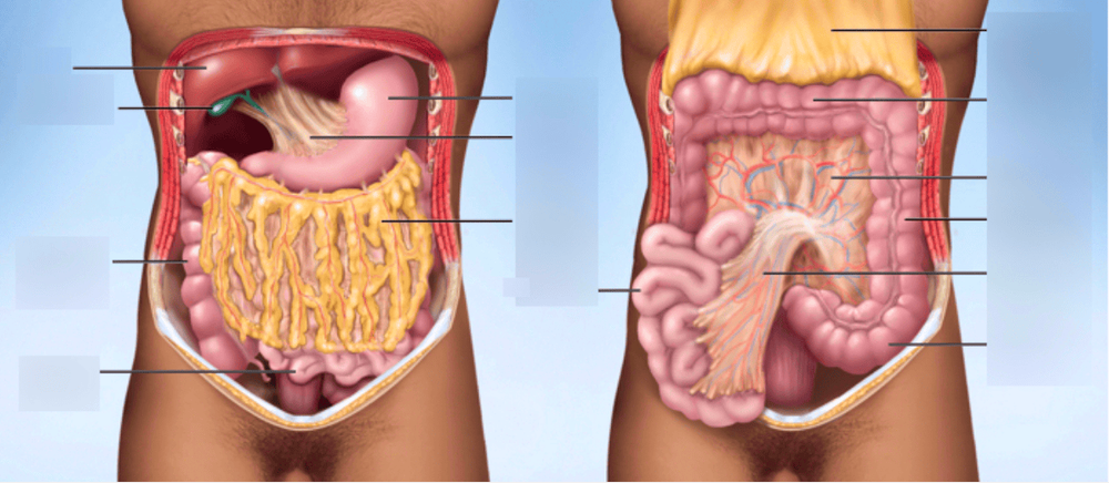
Mạc treo đại tràng và nạc treo ruột non
Mesenteric appendix : A triangular peritoneal fold surrounding the appendix, attached to the back of the lower end of the mesentery of the small intestine close to the ileocecal junction. It usually reaches the tip of the appendix but sometimes does not reach the external 1/3. It covers the blood vessels, nerves, and lymphatic vessels of the appendix.
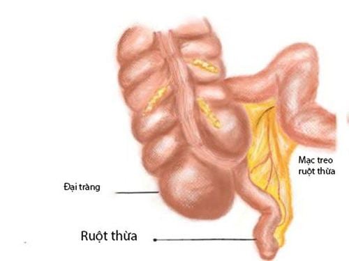
Hình ảnh mạc treo ruột thừa
Transverse mesentery: is a wide peritoneal fold connecting the transverse colon to the posterior abdominal wall, the two leaves of the transverse mesocolon passing from the anterior surface of the pancreatic head and the anterior border of the body of the pancreas to the posterior surface can be separated. from the great omentum. Posteriorly, the inferior leaflets cover the lower surface of the pancreas and run anteriorly to the transverse and ascending parts of the duodenum. In between the two leaves are the blood vessels, nerves, lymphatic vessels of the transverse colon. The sigmoid mesentery: This is a fold of peritoneum connecting the sigmoid colon to the iliac wall, its root attachment is an inverted V with a crest near the bifurcation of the left common iliac artery; The left stroke of the inverted V descends within the left major lumbar muscle and the right stroke runs into the pelvis to terminate at the midline at the level of the third sacral vertebra. The superior sigmoid and rectovaginal vessels run between its leaflets and the left ureter descends into the pelvis at the posterior apex of the V.


